 Back
BackCentral Nervous System: Structure, Development, and Organization
Study Guide - Smart Notes
 Tailored notes based on your materials, expanded with key definitions, examples, and context.
Central Nervous System (CNS) Overview
Definition and Components
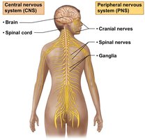
The central nervous system (CNS) is composed of the brain and spinal cord. It is responsible for integrating sensory information and responding accordingly. The CNS is distinct from the peripheral nervous system (PNS), which includes cranial nerves, spinal nerves, and ganglia.
Brain: The control center for processing and interpreting sensory information and directing responses.
Spinal Cord: Conducts signals to and from the brain and controls reflex activities.
Development of the Brain
Embryological Origins
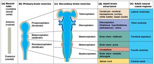
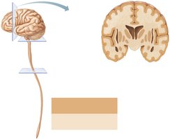
The brain and spinal cord originate from the neural tube during embryonic development. The anterior (rostral) end of the neural tube expands and forms three primary brain vesicles, which further differentiate into five secondary vesicles, eventually giving rise to the major structures of the adult brain.
Primary brain vesicles: Prosencephalon (forebrain), Mesencephalon (midbrain), Rhombencephalon (hindbrain)
Secondary brain vesicles: Telencephalon, Diencephalon, Mesencephalon, Metencephalon, Myelencephalon
Adult brain structures: Cerebrum, diencephalon, brain stem (midbrain, pons, medulla oblongata), cerebellum
Adult Brain Regions
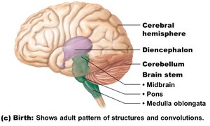
At birth, the brain exhibits a distinct pattern of structures and convolutions, including the cerebral hemispheres, diencephalon, cerebellum, and brain stem (midbrain, pons, medulla oblongata).
Organization of Gray and White Matter
Distribution Patterns
The CNS displays characteristic patterns of gray matter (neuron cell bodies, dendrites, glial cells) and white matter (myelinated axons):
In the spinal cord: central gray matter surrounded by white matter.
In the brain stem: additional gray matter nuclei embedded within white matter.
In the cerebrum and cerebellum: outer cortex of gray matter, inner white matter, and deep gray matter nuclei.
Ventricles of the Brain
Structure and Function
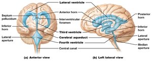
The brain contains interconnected, fluid-filled chambers called ventricles, which are continuous with the central canal of the spinal cord and filled with cerebrospinal fluid (CSF). They are lined by ependymal cells and serve to cushion and nourish the brain.
Lateral ventricles: Paired, C-shaped chambers in each hemisphere, separated by the septum pellucidum.
Third ventricle: Located in the diencephalon, connected to lateral ventricles via interventricular foramen.
Fourth ventricle: Located dorsal to the pons and medulla, connected to the third ventricle by the cerebral aqueduct; continuous with the central canal of the spinal cord.
Cerebral Hemispheres
Surface Features and Lobes
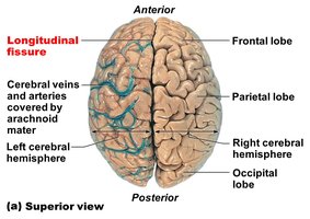
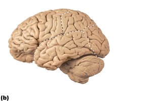
The cerebral hemispheres form the superior part of the brain and account for the majority of brain mass. The surface is marked by gyri (ridges), sulci (shallow grooves), and fissures (deep grooves). Major fissures include the longitudinal fissure (separates hemispheres) and the transverse cerebral fissure (separates cerebrum from cerebellum).
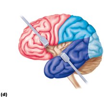
Each hemisphere is divided into five lobes: frontal, parietal, temporal, occipital, and insula.
Major sulci: central sulcus (separates frontal and parietal lobes), parieto-occipital sulcus, lateral sulcus (outlines temporal lobe).
Functional Areas of the Cerebral Cortex
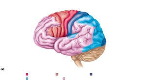
The cerebral cortex is the site of conscious mind activities, including awareness, sensory perception, voluntary motor initiation, communication, memory storage, and understanding. It is a thin (2–4 mm) layer of gray matter covering the cerebral hemispheres.
Three types of functional areas: motor (voluntary movement), sensory (conscious sensation), association (integration of information).
Each hemisphere controls the contralateral side of the body.
Lateralization: specialization of function in one hemisphere (e.g., language in the left hemisphere).
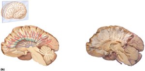
Motor and Sensory Maps
Specific regions of the cortex correspond to control of particular body parts, represented by the motor homunculus (precentral gyrus) and sensory homunculus (postcentral gyrus). These maps illustrate the proportional representation of body regions based on the density of motor or sensory innervation.
Cerebral White Matter and Basal Nuclei
Cerebral White Matter
White matter consists of myelinated fibers organized into tracts:
Association fibers: Connect regions within the same hemisphere.
Commissural fibers: Connect corresponding gray areas of the two hemispheres (e.g., corpus callosum).
Projection fibers: Connect the cortex with lower brain regions and the spinal cord.
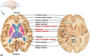
Basal Nuclei (Ganglia)
The basal nuclei are deep gray matter structures involved in the regulation of voluntary motor activities. They include the caudate nucleus, putamen, and globus pallidus. Disorders of the basal nuclei, such as Parkinson's disease and Huntington's disease, result in abnormal movements.
Diencephalon
Major Components
The diencephalon consists of three paired gray-matter structures: thalamus, hypothalamus, and epithalamus. These structures enclose the third ventricle and are involved in sensory relay, homeostatic regulation, and endocrine function.
Thalamus: Relay station for sensory and motor signals to the cerebral cortex.
Hypothalamus: Main visceral control center, regulating autonomic functions, emotions, body temperature, hunger, thirst, and the endocrine system.
Epithalamus: Contains the pineal gland, which secretes melatonin for sleep-wake regulation.
