Back
BackCentral Nervous System: Structure, Development, and Functional Organization
Study Guide - Smart Notes
 Tailored notes based on your materials, expanded with key definitions, examples, and context.
Central Nervous System (CNS)
Overview of the CNS
The central nervous system (CNS) is composed of the brain and spinal cord. It is responsible for integrating sensory information and coordinating bodily functions. Embryologically, the CNS develops from a structure called the neural tube.
Rostral: Toward the forehead (upper)
Caudal: Toward the spinal cord (lower)
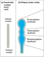
Embryonic Development of the Human Brain
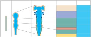
Neural Tube and Brain Vesicles
The brain and spinal cord originate from the neural tube. As development proceeds, the neural tube forms three primary brain vesicles, which further differentiate into secondary vesicles and adult brain structures.
Primary brain vesicles:
Prosencephalon (forebrain)
Mesencephalon (midbrain)
Rhombencephalon (hindbrain)
Secondary brain vesicles:
Telencephalon → Cerebrum
Diencephalon → Thalamus, hypothalamus, epithalamus, retina
Mesencephalon → Midbrain
Metencephalon → Pons, cerebellum
Myelencephalon → Medulla oblongata
Brain Regions and Organization
Major Brain Regions
The adult brain is organized into four main regions:
Cerebral hemispheres
Diencephalon
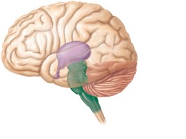
Brain stem (midbrain, pons, medulla oblongata)
Cerebellum
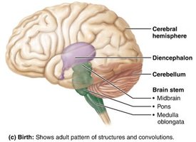
Structural Relationships
The brainstem connects the brain to the spinal cord and is composed of three main parts: the midbrain, pons, and medulla oblongata. The cerebellum lies posterior to the brainstem and is involved in motor coordination.
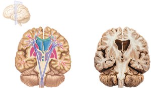
Internal Anatomy of the Brain
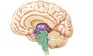
Midsagittal Section
A midsagittal section reveals the internal organization of the brain, including the cerebral cortex, white matter, basal nuclei, thalamus, hypothalamus, brainstem, and cerebellum.
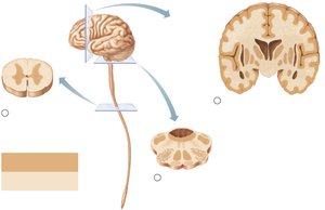
Gray Matter and White Matter in the CNS
Distribution Patterns
The CNS exhibits a basic pattern of gray matter (neuron cell bodies, nonmyelinated neurons) surrounded by white matter (myelinated and nonmyelinated axons). This pattern varies in different regions:
Spinal cord: Central gray matter surrounded by white matter
Brainstem: Additional gray matter nuclei within white matter
Cerebrum and cerebellum: Outer cortex of gray matter, inner white matter, and scattered gray matter nuclei
Cerebral Hemispheres
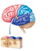
Surface Features and Lobes
The cerebral hemispheres form the superior part of the brain and account for about 83% of its mass. The surface is marked by gyri (ridges), sulci (shallow grooves), and fissures (deep grooves). Major lobes include:
Frontal lobe
Parietal lobe
Temporal lobe
Occipital lobe
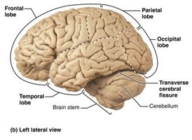
Longitudinal and Transverse Fissures
The longitudinal fissure separates the left and right cerebral hemispheres, while the transverse cerebral fissure separates the cerebrum from the cerebellum.
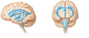
Ventricles of the Brain
Structure and Function
Ventricles are fluid-filled chambers within the brain that are continuous with each other and the central canal of the spinal cord. They are filled with cerebrospinal fluid (CSF) and lined by ependymal cells. The main ventricles include:
Lateral ventricles: Paired, C-shaped chambers in each hemisphere
Third ventricle: Located in the diencephalon
Fourth ventricle: Located in the hindbrain, continuous with the central canal
Ventricles are connected by the interventricular foramen and cerebral aqueduct.
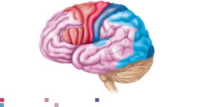
Cerebral Cortex
Functional Areas
The cerebral cortex is the site of conscious mind functions, including awareness, sensory perception, voluntary motor initiation, communication, memory storage, and understanding. It contains three main types of functional areas:
Motor areas: Control voluntary movements (located in the frontal lobe)
Sensory areas: Conscious awareness of sensation (parietal, temporal, occipital lobes)
Association areas: Integrate diverse information
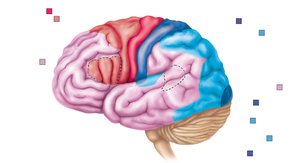
Motor Areas
Primary (somatic) motor cortex: Located in the precentral gyrus; initiates voluntary movement of skeletal muscle
Premotor cortex: Plans movements, controls learned and patterned motor skills
Broca’s area: Motor speech area, usually in the left hemisphere
Frontal eye field: Controls voluntary eye movements

Sensory Areas
Primary somatosensory cortex: Located in the postcentral gyrus; receives sensory information from skin, muscles, and joints
Somatosensory association cortex: Integrates sensory input for understanding
Primary visual cortex: Receives visual information from the retinas
Visual association area: Interprets visual stimuli
Primary auditory cortex: Interprets information from the inner ear
Auditory association area: Stores memories of sounds and permits perception of sound stimulus

Somatotopy and Body Maps
The body is represented spatially in the primary motor and somatosensory cortices, depicted by a homunculus. Areas with finer motor control or greater sensitivity have larger cortical representations.

Cerebral White Matter
Fiber Tracts
Cerebral white matter is responsible for communication within the brain and between the brain and spinal cord. It consists of myelinated fibers classified as:
Association fibers: Connect different parts of the same hemisphere
Commissural fibers: Connect gray matter of the two hemispheres (e.g., corpus callosum)
Projection fibers: Connect the cortex with lower brain regions or the spinal cord
Ascending and Descending Pathways
Neuronal Pathways
Major spinal tracts are organized into multi-neuron pathways with the following features:
Decussation: Most pathways cross from one side of the CNS to the other
Relay: Consist of two or three neurons in sequence
Somatotopy: Spatial correspondence between CNS regions and body regions
Symmetry: Pathways are paired symmetrically
Ascending Pathways
Dorsal column–medial lemniscal pathways: Discriminative touch and vibration
Spinothalamic pathways: Pain, temperature, coarse touch, and pressure
Spinocerebellar tracts: Muscle or tendon stretch to cerebellum for coordination
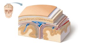
Meninges of the CNS
Protective Membranes
The meninges are three connective tissue membranes that cover and protect the CNS:
Dura mater: Strongest, outermost layer; forms dural septa and venous sinuses
Arachnoid mater: Middle layer with web-like extensions; contains CSF and blood vessels
Pia mater: Delicate, innermost layer; clings tightly to the brain and contains blood vessels
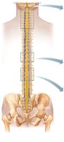
Spinal Cord
Structure and Function
The spinal cord is enclosed within the vertebral column and extends from the foramen magnum to the lumbar region. It provides two-way communication between the brain and body and mediates spinal reflexes.