 Back
BackCentral Nervous System: Structure, Development, and Functional Organization
Study Guide - Smart Notes
 Tailored notes based on your materials, expanded with key definitions, examples, and context.
Central Nervous System (CNS): Overview
Major Functions and Structures
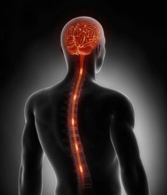
The Central Nervous System (CNS) consists of the brain and spinal cord. It is responsible for integrating sensory information, planning and monitoring movement, maintaining homeostasis, and supporting higher mental functions such as language and learning. The Peripheral Nervous System (PNS) connects the CNS to limbs and organs, facilitating sensory and motor functions.
Interpretation of sensory information: Processing input from sensory organs.
Planning & monitoring movement: Initiating and coordinating voluntary and involuntary movements.
Maintaining homeostasis: Regulating internal environment (e.g., temperature, pH).
Higher mental functions: Language, learning, memory, and reasoning.
Development of the Human Brain
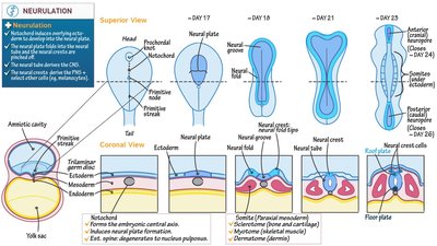
Neurulation and Neural Tube Formation
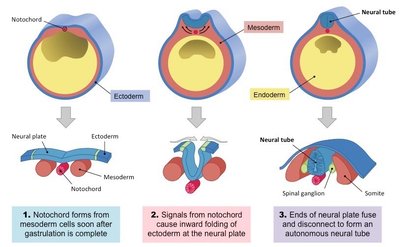
The CNS develops from the ectodermal layer of the embryo through a process called neurulation. This process forms the neural plate, which folds to create the neural tube—the precursor to the brain and spinal cord.
Neural plate: Thickened region of ectoderm that gives rise to the CNS.
Neural groove and folds: The neural plate invaginates, forming a groove flanked by neural folds.
Neural tube: The folds fuse to form the neural tube, which sinks deeper into the embryo.
Neural crest cells: Migrate to form peripheral nervous system structures, including dorsal root ganglia.
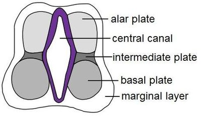
Neural Tube Differentiation
Within the neural tube, two main clusters of neuroblast cells differentiate:
Dorsal alar plate: Becomes interneurons; axons form white matter of the spinal cord.
Basal plate: Becomes motor neurons.
Neural crest cells: Form dorsal root ganglia and other PNS components.
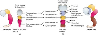
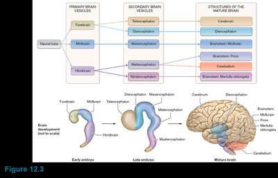
Brain Vesicle Formation and Regionalization
The neural tube expands and constricts to form three primary brain vesicles, which further subdivide into five secondary vesicles, each giving rise to specific adult brain structures.
Primary brain vesicles:
Prosencephalon (forebrain)
Mesencephalon (midbrain)
Rhombencephalon (hindbrain)
Secondary brain vesicles:
Telencephalon (cerebral hemispheres)
Diencephalon (thalamus, hypothalamus, epithalamus, retina)
Mesencephalon (midbrain)
Metencephalon (pons, cerebellum)
Myelencephalon (medulla oblongata)
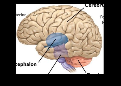
Structural Organization of the Brain
Major Brain Regions
The mature brain is organized into four main regions:
Cerebrum: Largest part, responsible for higher mental functions.
Diencephalon: Processes and relays information, maintains homeostasis.
Brainstem: Controls basic life functions and relays information between brain and spinal cord.
Cerebellum: Coordinates movement and balance.
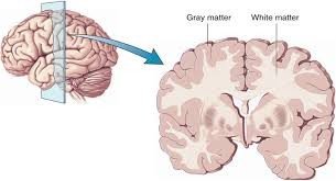
Gray Matter and White Matter
The brain and spinal cord are composed of gray matter (neuronal cell bodies, dendrites) and white matter (myelinated axons). Their arrangement varies by region:
Gray matter: Outer cortex of the brain, inner core of the spinal cord.
White matter: Inner regions of the brain, outer regions of the spinal cord.
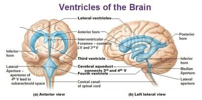
Ventricular System of the Brain
Ventricles and Cerebrospinal Fluid (CSF)
The brain contains interconnected, fluid-filled cavities called ventricles that produce and circulate cerebrospinal fluid (CSF). CSF cushions the brain, removes waste, and provides nutrients.
Lateral ventricles: Paired, located in cerebral hemispheres, separated by septum pellucidum.
Third ventricle: Located in the diencephalon, connected to lateral ventricles via interventricular foramen.
Fourth ventricle: Located in the hindbrain, connected to third ventricle via cerebral aqueduct; opens to subarachnoid space via lateral and median apertures.

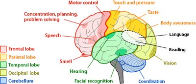
Cerebral Cortex and Functional Areas
Lobes of the Cerebral Cortex
The cerebral cortex is divided into five lobes per hemisphere, each with specialized functions:
Frontal lobe: Planning, thinking, behavior, conscience, personality.
Parietal lobe: Sensory integration, attention.
Temporal lobe: Hearing, language, memory, emotions.
Occipital lobe: Visual information processing.
Insular lobe: Deep within the lateral sulcus, involved in taste and visceral sensation.

Gyri, Sulci, and Fissures
The surface of the cerebral cortex is highly folded, increasing surface area:
Gyri: Elevated ridges (e.g., precentral and postcentral gyri).
Sulci: Shallow grooves (e.g., central sulcus, parieto-occipital sulcus, lateral sulcus).
Fissures: Deep grooves (e.g., longitudinal fissure, transverse cerebral fissure).
Functional Areas of the Cortex
The cortex contains distinct areas for motor, sensory, and association functions:
Motor areas: Control voluntary movement (e.g., primary motor cortex in precentral gyrus).
Sensory areas: Receive and process sensory input (e.g., primary somatosensory cortex in postcentral gyrus, visual cortex in occipital lobe, auditory cortex in temporal lobe).
Association areas: Integrate information and support higher functions (e.g., prefrontal cortex, limbic association area).
White Matter and Basal Nuclei
Cerebral White Matter
White matter consists of myelinated axons organized into tracts:
Commissural fibers: Connect right and left hemispheres (e.g., corpus callosum).
Projection fibers: Connect cortex with lower brain regions and spinal cord.
Association fibers: Connect different parts of the same hemisphere.
Basal Nuclei
The basal nuclei are clusters of gray matter deep within the cerebral hemispheres. They are involved in movement regulation, behavior, cognition, and perception. The main components are:
Caudate nucleus
Putamen
Globus pallidus
Corpus striatum: Caudate nucleus + putamen
Summary Table: Brain Vesicle Development
Primary Vesicle | Secondary Vesicle | Adult Brain Structure |
|---|---|---|
Prosencephalon (Forebrain) | Telencephalon | Cerebral hemispheres (cerebrum) |
Prosencephalon (Forebrain) | Diencephalon | Thalamus, hypothalamus, epithalamus, retina |
Mesencephalon (Midbrain) | Mesencephalon | Midbrain |
Rhombencephalon (Hindbrain) | Metencephalon | Pons, cerebellum |
Rhombencephalon (Hindbrain) | Myelencephalon | Medulla oblongata |
Key Terms and Concepts
Neurulation: Formation of the neural tube from the neural plate.
Neural crest: Cells that migrate to form the PNS and other structures.
Gray matter: Regions of the CNS containing neuron cell bodies and dendrites.
White matter: Regions of the CNS containing myelinated axons.
Basal nuclei: Deep brain structures involved in movement and cognition.
Ventricles: Fluid-filled cavities in the brain that produce and circulate CSF.
