 Back
BackCentral Nervous System: Structure, Development, and Functional Organization
Study Guide - Smart Notes
 Tailored notes based on your materials, expanded with key definitions, examples, and context.
Central Nervous System Overview
Definition and Components
The Central Nervous System (CNS) is composed of the brain and spinal cord. It is responsible for integrating sensory information and coordinating bodily functions. Embryologically, the CNS originates from the neural tube, which develops into the brain and spinal cord.
Rostral: Refers to structures toward the forehead (upper).
Caudal: Refers to structures toward the spinal cord (lower).
Embryonic Development of the Brain
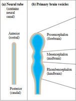
Neural Tube and Brain Vesicles
The neural tube forms three primary brain vesicles:
Prosencephalon: Forebrain
Mesencephalon: Midbrain
Rhombencephalon: Hindbrain
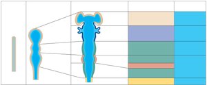
These primary vesicles further differentiate into secondary brain vesicles, which give rise to adult brain structures:
Telencephalon: Cerebral hemispheres
Diencephalon: Thalamus, hypothalamus, epithalamus, retina
Mesencephalon: Midbrain
Metencephalon: Pons and cerebellum
Myelencephalon: Medulla oblongata
Brain Regions and Organization
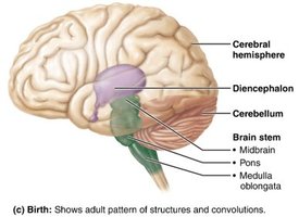
Major Regions of the Adult Brain
The adult brain is organized into four main regions:
Cerebral Hemispheres
Diencephalon
Brain Stem: Midbrain, pons, medulla oblongata
Cerebellum
Structural Features
The brainstem consists of three prominent structures: midbrain, pons (largest bump), and medulla. The spinal cord continues from the brainstem through the foramen magnum.
Gray Matter and White Matter in the CNS
Distribution Patterns
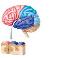
The CNS exhibits a basic pattern of central cavity surrounded by gray matter, with white matter external to gray matter. This pattern changes as one ascends from the spinal cord to the brainstem and cerebrum.
Gray Matter: Contains neuron cell bodies and short, nonmyelinated neurons.
White Matter: Composed mostly of myelinated axons, with some nonmyelinated axons.
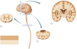
In the cerebrum and cerebellum, an outer layer of gray matter called the cortex is present, along with scattered gray matter nuclei within white matter.
Cerebral Hemispheres
Surface Markings and Lobes
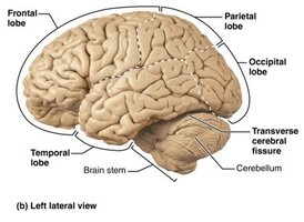
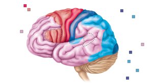
The cerebral hemispheres form the superior part of the brain and account for 83% of its mass. Surface features include:
Gyri: Ridges (folds)
Sulci: Shallow grooves
Fissures: Deep grooves
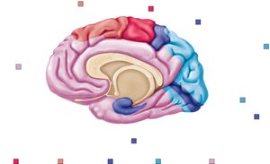
Each hemisphere is divided into five lobes: frontal, parietal, temporal, occipital, and insula (not shown in all diagrams). The hemispheres are separated by the longitudinal fissure and connected by the corpus callosum.
Functional Regions
Cerebral Cortex: Thin outer region of gray matter
Cerebral White Matter: Deeper region, appears white
Ventricles of the Brain
Structure and Function
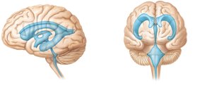
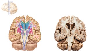
Brain ventricles are fluid-filled chambers lined by ependymal cells and filled with cerebrospinal fluid (CSF). They are continuous with each other and the central canal of the spinal cord.
Lateral Ventricles: Paired, C-shaped chambers in each hemisphere
Third Ventricle: Located in the diencephalon
Fourth Ventricle: Located in the hindbrain, continuous with the central canal
Cerebral Cortex Functional Areas
Motor Areas
Motor areas are located in the frontal lobe and control voluntary movement:
Primary (Somatic) Motor Cortex: Located in the precentral gyrus; initiates voluntary movement of skeletal muscle.
Premotor Cortex: Plans movements and controls learned, patterned motor skills.
Broca’s Area: Motor speech area, usually in the left hemisphere.
Frontal Eye Field: Controls voluntary eye movements.
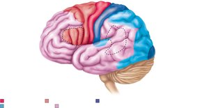
Sensory Areas
Sensory areas are located in the parietal, temporal, and occipital lobes. They include:
Primary Somatosensory Cortex: Located in the postcentral gyrus; receives sensory information from skin, muscles, and joints.
Visual Cortex: Located in the occipital lobe; receives and interprets visual information.
Auditory Cortex: Located in the temporal lobe; interprets sound information.
Lateralization of Cortical Functioning
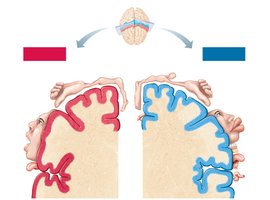
Lateralization refers to the division of labor between the hemispheres:
Left Hemisphere: Language, math, logic
Right Hemisphere: Visual-spatial skills, intuition, emotion, artistic and musical skills
Hemispheres communicate via fiber tracts and functional integration. Cerebral dominance refers to the hemisphere dominant for language (usually left).
Cerebral White Matter
Fiber Tracts
Cerebral white matter is responsible for communication within the brain and between the brain and spinal cord. Fiber tracts are classified by direction:
Association Fibers: Connect different parts of the same hemisphere
Commissural Fibers: Connect gray matter of the two hemispheres (e.g., corpus callosum)
Projection Fibers: Connect hemispheres with lower brain or spinal cord
Somatotopy and Body Maps
Homunculus Representation
The body is spatially represented in the primary motor cortex (precentral gyrus) and primary somatosensory cortex (postcentral gyrus). This mapping is depicted by the homunculus, which shows the relative size of cortical areas dedicated to different body regions.
Ascending and Descending Pathways
Spinal Tracts and Pathways
Major spinal tracts are part of multi-neuron pathways:
Decussation: Most pathways cross from one side of the CNS to the other.
Relay: Pathways consist of chains of two or three neurons.
Somatotopy: Spatial relationships in the CNS correspond to those in the body.
Symmetry: Pathways are paired symmetrically.
Ascending Pathways
Dorsal Column–Medial Lemniscal Pathways: Discriminative touch and vibration
Spinothalamic Pathways: Pain, temperature, coarse touch, pressure
Spinocerebellar Tracts: Muscle or tendon stretch, coordination
Meninges of the CNS
Dura Mater
The dura mater is the strongest meninx, consisting of two layers in the brain (periosteal and meningeal). It forms partitions called dural septa, which limit excessive movement of the brain.
Falx Cerebri: In longitudinal fissure
Falx Cerebelli: Along vermis of cerebellum
Tentorium Cerebelli: Over cerebellum and in transverse fissure
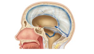
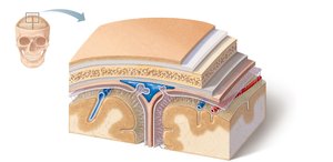
Arachnoid Mater
The arachnoid mater is the middle meningeal layer, separated from the dura mater by the subdural space. The subarachnoid space contains CSF and blood vessels. Arachnoid granulations allow CSF reabsorption into venous blood.
Pia Mater
The pia mater is a delicate layer that clings tightly to the brain, following every convolution and containing many blood vessels.
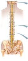
Spinal Cord
Structure and Function
The spinal cord is enclosed in the vertebral column and begins at the foramen magnum. It provides two-way communication between the brain and body and carries out spinal reflexes.
Summary Table: Major Brain Regions and Their Functions
Region | Main Function |
|---|---|
Cerebral Hemispheres | Conscious mind, voluntary movement, sensory perception |
Diencephalon | Relay and processing of sensory information, homeostasis |
Brain Stem | Basic life functions, pathway for information between brain and body |
Cerebellum | Coordination of movement, balance |
Additional info: The notes include expanded academic context and definitions to ensure completeness and clarity for exam preparation.
