 Back
BackCentral Nervous System: Structure, Development, and Functional Organization
Study Guide - Smart Notes
 Tailored notes based on your materials, expanded with key definitions, examples, and context.
The Central Nervous System (CNS)
Overview and Importance
The central nervous system (CNS) comprises the brain and spinal cord, serving as the primary control center for the body. Understanding its structure and function is essential for diagnosing and treating neurological injuries and disorders.
Brain Development
Embryological Formation
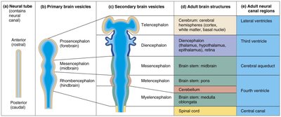
During embryonic development, the CNS originates from the neural tube. The anterior end of the neural tube expands and forms three primary brain vesicles:
Prosencephalon (forebrain)
Mesencephalon (midbrain)
Rhombencephalon (hindbrain)
These primary vesicles further differentiate into five secondary brain vesicles, which give rise to the major adult brain structures.
Primary Vesicle | Secondary Vesicle | Adult Structure | Neural Canal Region |
|---|---|---|---|
Prosencephalon | Telencephalon | Cerebrum (cerebral hemispheres) | Lateral ventricles |
Prosencephalon | Diencephalon | Thalamus, hypothalamus, epithalamus, retina | Third ventricle |
Mesencephalon | Mesencephalon | Midbrain | Cerebral aqueduct |
Rhombencephalon | Metencephalon | Pons, cerebellum | Fourth ventricle |
Rhombencephalon | Myelencephalon | Medulla oblongata | Fourth ventricle |
Spinal cord | - | Spinal cord | Central canal |
Brain Folding and Growth
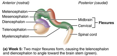
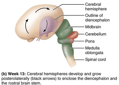
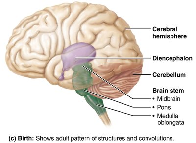
As the brain grows, it folds to fit within the skull. The telencephalon and diencephalon angle toward the brain stem, and cerebral hemispheres envelop the diencephalon and midbrain, increasing surface area.
Brain Regions and Organization
Major Regions
The adult brain is organized into four main regions:
Cerebral hemispheres
Diencephalon
Brain stem (midbrain, pons, medulla oblongata)
Cerebellum
Gray and White Matter Distribution
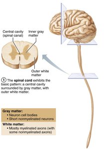
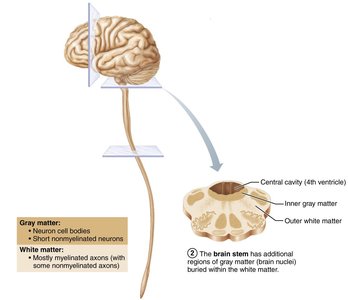
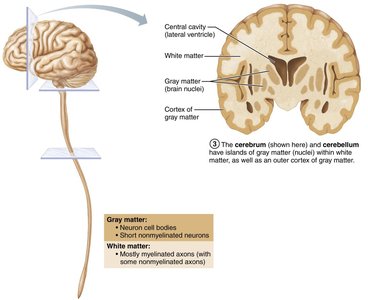
Gray matter consists of neuron cell bodies and nonmyelinated neurons, while white matter is composed of myelinated axons. The basic pattern is a central cavity surrounded by gray matter, with white matter external to it. This pattern changes in the brain stem and cerebrum, where additional nuclei and cortex are present.
Ventricles of the Brain
Structure and Function
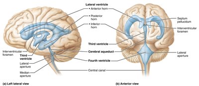
Brain ventricles are fluid-filled chambers lined by ependymal cells and filled with cerebrospinal fluid (CSF). The paired lateral ventricles are separated by the septum pellucidum and connect to the third ventricle via the interventricular foramen. The third ventricle connects to the fourth ventricle via the cerebral aqueduct. The fourth ventricle is continuous with the central canal of the spinal cord and connects to the subarachnoid space via three apertures.
Cerebral Hemispheres
Surface Markings and Lobes
The cerebral hemispheres account for 83% of brain mass and are marked by gyri (ridges), sulci (shallow grooves), and fissures (deep grooves). The longitudinal fissure separates the hemispheres, while the transverse cerebral fissure separates the cerebrum from the cerebellum. Each hemisphere is divided into five lobes: frontal, parietal, temporal, occipital, and insula.
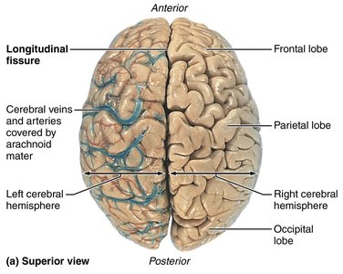
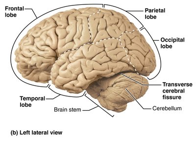
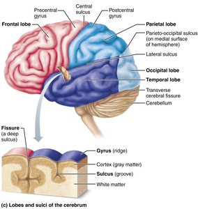
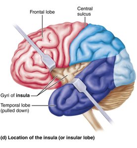
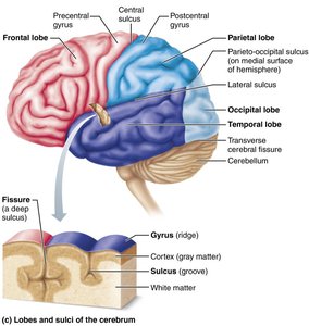
Major Sulci and Regions
Central sulcus: separates precentral gyrus (frontal lobe) and postcentral gyrus (parietal lobe)
Parieto-occipital sulcus: separates occipital and parietal lobes
Lateral sulcus: outlines temporal lobes
Each hemisphere contains three basic regions: the superficial gray matter cortex, internal white matter, and deep basal nuclei.
Cerebral Cortex
Functional Areas
The cerebral cortex is the site of conscious mind, including awareness, sensory perception, voluntary motor initiation, communication, memory, and understanding. It contains three types of functional areas:
Motor areas: control voluntary movement
Sensory areas: conscious awareness of sensation
Association areas: integrate diverse information
Each hemisphere controls the contralateral side of the body, and lateralization of function can occur.
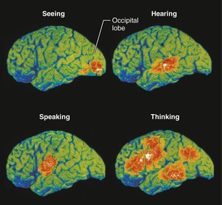
Motor Areas
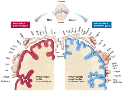
Primary motor cortex: located in precentral gyrus; controls precise, skilled movements
Premotor cortex: plans movements and coordinates actions
Broca’s area: motor speech area
Frontal eye field: controls voluntary eye movements
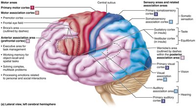
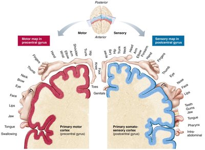
Sensory Areas
Primary somatosensory cortex: receives sensory information from skin and proprioceptors
Somatosensory association cortex: integrates sensory input for object recognition
Visual areas: interpret visual information
Auditory areas: interpret sound
Vestibular cortex: balance awareness
Olfactory cortex: smell perception
Gustatory cortex: taste perception
Visceral sensory area: perception of visceral sensations
Association Areas
Anterior association area (prefrontal cortex): intellect, cognition, personality
Posterior association area: pattern and face recognition, spatial localization, language comprehension
Limbic association area: emotional impact and memory formation
Lateralization of Function
Left hemisphere: language, math, logic
Right hemisphere: visual-spatial skills, intuition, emotion, artistic abilities
Cerebral White Matter
Fiber Types
Association fibers: connect different parts of the same hemisphere
Commissural fibers: connect gray matter of two hemispheres
Projection fibers: connect hemispheres with lower brain or spinal cord
Basal Nuclei
Structure and Function
Basal nuclei (ganglia) include the caudate nucleus, putamen, and globus pallidus. They influence muscle movements, regulate intensity of movements, filter out inappropriate responses, and inhibit unnecessary movements. Disorders include Parkinson’s and Huntington’s disease.
Diencephalon
Components
Thalamus: relay station for sensory and motor information
Hypothalamus: main visceral control center; regulates homeostasis, autonomic functions, endocrine system
Epithalamus: contains pineal gland, regulates sleep-wake cycle
Brain Stem
Regions and Functions
Midbrain: visual and auditory reflex centers, motor control
Pons: relays information, controls respiratory rhythm
Medulla oblongata: autonomic reflex center, cardiovascular and respiratory regulation
Cerebellum
Structure and Function
The cerebellum coordinates voluntary movements, balance, and posture. It processes input from the cortex, brain stem, and sensory receptors, and sends instructions for smooth, coordinated muscle activity.
Functional Brain Systems
Limbic System
Mediates emotional responses and memory processing; includes structures such as the amygdaloid body, hippocampus, and cingulate gyrus.
Reticular Formation
Maintains cerebral cortical alertness, filters repetitive stimuli, and regulates skeletal and visceral muscle activity.
Protection of the Brain
Meninges
Dura mater: tough, outer layer
Arachnoid mater: middle, web-like layer
Pia mater: delicate, inner layer
Cerebrospinal Fluid (CSF)
CSF cushions the brain, provides buoyancy, removes waste, and nourishes neural tissue. It is produced by the choroid plexus and circulates through ventricles and subarachnoid space.
Blood Brain Barrier
The blood brain barrier protects the brain from harmful substances by regulating passage through endothelial cells and tight junctions.
Spinal Cord
Structure and Function
The spinal cord provides two-way communication between the brain and body and serves as a major reflex center. It is protected by bone, meninges, and CSF, and terminates at the conus medullaris.
Cross-Sectional Anatomy
Gray matter: dorsal, ventral, and lateral horns
White matter: ascending, descending, and transverse tracts
Neuronal Pathways
Ascending (Sensory) Pathways
Dorsal column–medial lemniscal: discriminative touch, pressure, proprioception
Spinothalamic: pain, temperature, coarse touch
Spinocerebellar: subconscious proprioception
Descending (Motor) Pathways
Direct (pyramidal): fast, skilled movements
Indirect: balance, posture, coarse movements, head/eye coordination
Clinical Considerations
Traumatic brain injuries: concussion, contusion, hemorrhage, edema
Cerebrovascular accidents (stroke): ischemia, hemiplegia, TIA
Degenerative disorders: Alzheimer’s, Parkinson’s, Huntington’s
Spinal cord trauma: paralysis, transection, poliomyelitis, ALS
Developmental disorders: cerebral palsy, anencephaly, spina bifida
