 Back
BackLecture - Ch 13 Central Nervous System
Study Guide - Smart Notes
 Tailored notes based on your materials, expanded with key definitions, examples, and context.
The Central Nervous System (CNS)
Overview and Cephalization
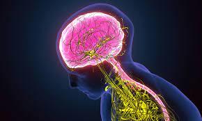
The central nervous system (CNS) consists of the brain and spinal cord, serving as the main control center for the body. Cephalization refers to the evolutionary development of the anterior portion of the CNS, resulting in an increased number of neurons in the head and the highest level of complexity in the human brain.
Embryonic Development of the CNS
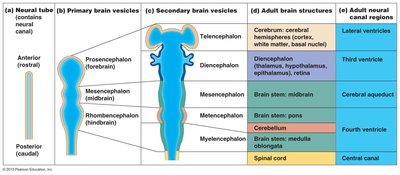
The brain and spinal cord originate from the neural tube during embryonic development. The anterior end of the neural tube forms three primary brain vesicles: the prosencephalon (forebrain), mesencephalon (midbrain), and rhombencephalon (hindbrain). These primary vesicles further differentiate into five secondary brain vesicles, which give rise to the major adult brain structures.
Telencephalon: Forms the cerebral hemispheres.
Diencephalon: Forms the epithalamus, thalamus, hypothalamus, and retina.
Mesencephalon: Remains as the midbrain.
Metencephalon: Forms the pons and cerebellum.
Myelencephalon: Forms the medulla oblongata.
Adult Brain Regions
Major Regions and Structures
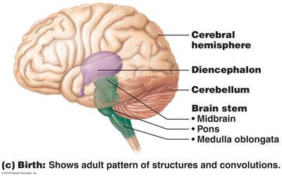
The adult brain is organized into four main regions: cerebral hemispheres, diencephalon, brain stem (midbrain, pons, medulla), and cerebellum. Each region has distinct functions and anatomical features.
Cerebral hemispheres: Responsible for higher cognitive functions.
Diencephalon: Includes thalamus, hypothalamus, and epithalamus.
Brain stem: Controls vital functions and connects the brain to the spinal cord.
Cerebellum: Coordinates movement and balance.
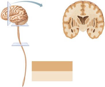
Gray and White Matter in the CNS
Distribution and Function
Gray matter consists of neuron cell bodies and short nonmyelinated neurons, while white matter is composed mainly of myelinated axons. The cerebrum and cerebellum contain islands of gray matter (nuclei) within white matter, as well as an outer cortex of gray matter.
Gray matter: Site of synaptic integration and processing.
White matter: Facilitates communication between different CNS regions.
Cerebral Hemispheres
Surface Markings and Lobes
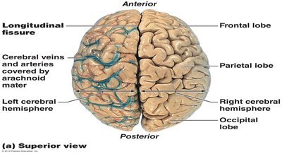
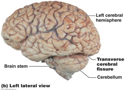
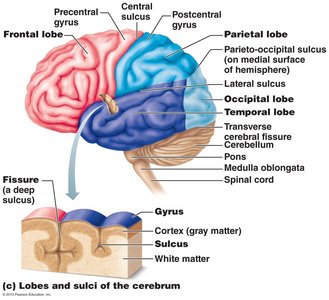
The cerebral hemispheres form the superior part of the brain and account for 83% of its mass. Surface markings include gyri (ridges), sulci (shallow grooves), and fissures (deep grooves). The longitudinal fissure separates the two hemispheres, while the transverse cerebral fissure separates the cerebrum from the cerebellum.
Lobes of the Cerebrum
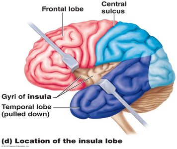
Each hemisphere is divided into five lobes: frontal, parietal, temporal, occipital, and insula. The first four are named after the overlying skull bones, while the insular lobe is buried under portions of the temporal, parietal, and frontal lobes.

Cerebral Cortex
Functional Areas
The cerebral cortex is the site of conscious mind, including awareness, sensory perception, voluntary motor initiation, communication, memory storage, and understanding. It contains three types of functional areas:
Motor areas: Control voluntary movement.
Sensory areas: Provide conscious awareness of sensation.
Association areas: Integrate diverse information.
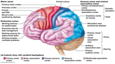
Motor Areas
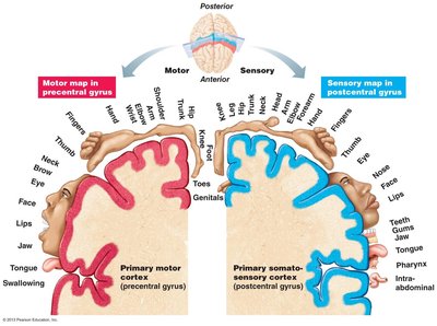
Motor areas are located in the frontal lobe and include the primary motor cortex (precentral gyrus), premotor cortex, Broca’s area, and frontal eye field. The primary motor cortex contains large pyramidal cells that allow conscious control of precise, skilled skeletal muscle movements.


Motor Homunculus
The motor homunculus is a spatial map of the body in the primary motor cortex, illustrating the contralateral motor innervation of body regions. Areas with the most precise motor control, such as the face, tongue, and hands, are disproportionately large in the homunculus.
Sensory Areas
Sensory areas are located in the parietal, insular, temporal, and occipital lobes. The primary somatosensory cortex receives general sensory information from the skin and proprioceptors, enabling spatial discrimination.

Association Areas
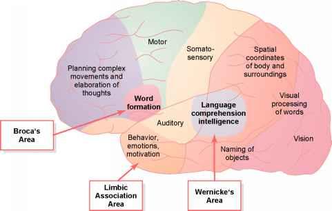
Association areas integrate sensory input for understanding objects, interpret visual stimuli, and store memories of sounds. The prefrontal cortex is involved in intellect, cognition, recall, and personality, while the posterior association area plays a role in recognizing patterns and faces and understanding language.
Lateralization and Dominance
Lateralization refers to the division of labor between hemispheres. The left hemisphere is dominant for language, math, and logic, while the right hemisphere specializes in visual-spatial skills, intuition, emotion, and artistic abilities.
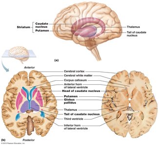
Cerebral White Matter and Basal Nuclei
White Matter Fiber Tracts
Cerebral white matter consists of myelinated fibers bundled into association, commissural, and projection tracts. These tracts facilitate communication within and between hemispheres and with lower CNS regions.
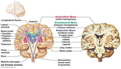
Basal Nuclei
Basal nuclei are deep gray matter structures involved in motor control, cognition, and emotion. They regulate the intensity of movements, filter out inappropriate responses, and inhibit unnecessary movements. Disorders such as Parkinson’s and Huntington’s disease are associated with basal nuclei dysfunction.
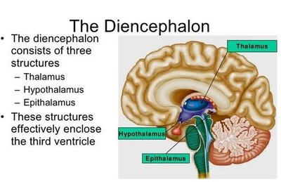
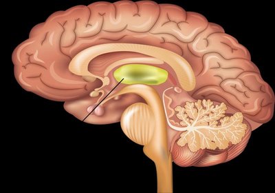
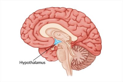
The Diencephalon
Thalamus, Hypothalamus, and Epithalamus
The diencephalon consists of the thalamus, hypothalamus, and epithalamus, which enclose the third ventricle. The thalamus acts as a relay station for sensory and motor information, the hypothalamus regulates homeostasis and endocrine functions, and the epithalamus contains the pineal gland, which secretes melatonin.
Brain Stem and Cerebellum
Brain Stem
The brain stem consists of the midbrain, pons, and medulla oblongata. It controls automatic behaviors necessary for survival and connects higher and lower neural centers. The medulla oblongata is an autonomic reflex center regulating cardiovascular and respiratory functions.
Cerebellum
The cerebellum coordinates precise, smooth movements and maintains balance. It processes input from the cortex, brain stem, and sensory receptors, and plays a role in cognitive functions such as thinking and language.
Functional Brain Systems
Limbic System and Reticular Formation
The limbic system is involved in emotional responses and memory formation, while the reticular formation governs brain arousal and consciousness. The reticular activating system (RAS) filters sensory input and maintains alertness.
Higher Mental Functions
Language, Memory, and EEG
Language is managed by Broca’s area (speech production) and Wernicke’s area (language comprehension). Memory involves the hippocampus and temporal lobe structures. Brain waves, recorded by EEG, reflect electrical activity and are used to diagnose disorders such as epilepsy.
Protection of the Brain
Meninges and Cerebrospinal Fluid (CSF)
The brain is protected by three layers of meninges: dura mater, arachnoid mater, and pia mater. CSF forms a liquid cushion, reducing brain weight and protecting against trauma. The blood-brain barrier maintains a stable environment by restricting entry of harmful substances.
Spinal Cord
Gross Anatomy and Protection
The spinal cord is enclosed in the vertebral column and protected by bone, meninges, and CSF. It provides two-way communication between the brain and body and serves as a major reflex center.
Cross-Sectional Anatomy
The spinal cord exhibits a central cavity surrounded by gray matter, with external white matter composed of myelinated fiber tracts. Gray matter is organized into dorsal, ventral, and lateral horns, each with specific functions.
Clinical Considerations
Brain and Spinal Cord Disorders
Traumatic brain injuries: Concussion, contusion, hemorrhage, and cerebral edema.
Cerebrovascular accidents (strokes): Ischemia and hemiplegia.
Degenerative disorders: Alzheimer’s, Parkinson’s, Huntington’s disease.
Spinal cord trauma: Paralysis, paresthesias, and transection injuries.
Congenital disorders: Anencephaly, cerebral palsy, spina bifida.
Developmental Aspects
Influences and Aging
Gender-specific areas develop in the CNS based on fetal testosterone. Maternal exposure to radiation, drugs, or infection can harm CNS development. Aging leads to cognitive decline and brain shrinkage, with excessive alcohol use and trauma causing signs of senility.
