 Back
BackCentral Nervous System: Structure, Function, and Development
Study Guide - Smart Notes
 Tailored notes based on your materials, expanded with key definitions, examples, and context.
Central Nervous System (CNS) & Brain Development
Overview of the CNS
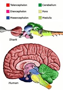
The central nervous system (CNS) is composed of the brain and spinal cord, serving as the primary control center for the body. Cephalization refers to the evolutionary development of the anterior portion of the CNS, resulting in the concentration of neural structures in the head.
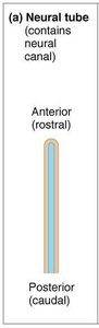
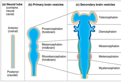
Brain and spinal cord originate embryologically from the neural tube.
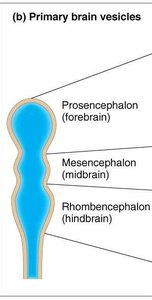
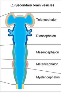
The anterior end of the neural tube expands and forms three primary brain vesicles, which further develop into five secondary vesicles.
The posterior end of the neural tube becomes the spinal cord.
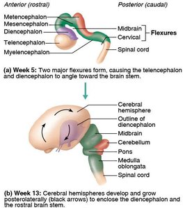
Brain Growth and Folding
The brain grows faster than the surrounding skull, resulting in folding to maximize surface area.
The forebrain moves toward the brain stem.
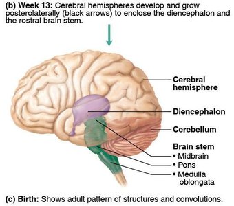
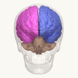
Cerebral hemispheres envelop the diencephalon and midbrain, increasing surface area through creasing and folding.
Brain Regions and Organization
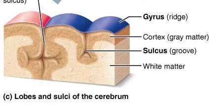
Gray Matter and White Matter
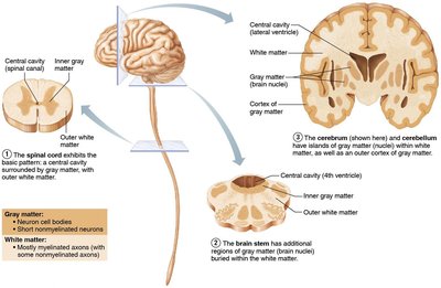
The brain and spinal cord are organized into gray and white matter.
Gray matter: Contains neuron cell bodies, dendrites, glial cells, and blood vessels; responsible for processing information.
White matter: Composed of myelinated and nonmyelinated axons; responsible for communication between regions.
Central cavity is surrounded by gray matter, with white matter external to it.
Brain stem contains additional gray matter nuclei within white matter.
Cerebral hemispheres and cerebellum have an outer layer of gray matter called the cortex.
Major Brain Regions
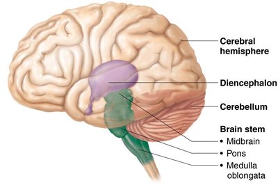
The adult brain is divided into four main regions:
Cerebral hemispheres
Diencephalon
Brain stem (midbrain, pons, medulla oblongata)
Cerebellum

Cerebral Hemispheres
Structure of the Cerebral Hemispheres
Each cerebral hemisphere consists of three basic regions:
Superficial cerebral cortex (gray matter)
Internal white matter
Basal nuclei (deep gray matter)

Cerebral Cortex
The cerebral cortex is the site of conscious mind, responsible for awareness, sensory perception, voluntary motor initiation, communication, memory storage, and understanding.
Composed of neuron cell bodies, dendrites, glial cells, and blood vessels; lacks axons.
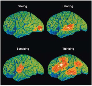
Functional imaging (PET, MRI) shows specific motor and sensory functions are localized in discrete cortical areas called domains.
Higher functions are distributed across multiple areas.

Functional Areas of the Cerebral Cortex
Four general considerations:
Three types of functional areas:
Motor areas: Control voluntary movement
Sensory areas: Conscious awareness of sensation
Association areas: Integrate diverse information
Each hemisphere controls the contralateral (opposite) side of the body.
Lateralization of cortical function can occur in only one hemisphere.
Conscious behavior involves the entire cortex.

Motor Areas
Primary (somatic) motor cortex: Contains pyramidal cells for conscious control of precise, skilled skeletal muscle movements. Motor homunculi represent contralateral motor innervation.
Premotor cortex: Plans movements, controls learned, repetitious, or patterned motor skills, and voluntary actions dependent on sensory feedback.
Broca’s area: Motor speech area directing muscles of speech production; active in planning speech and voluntary motor activities.
Frontal eye field: Controls voluntary eye movements.
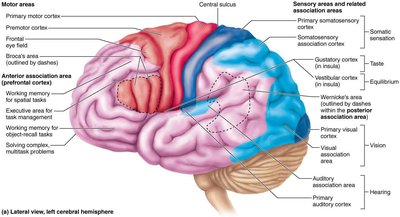
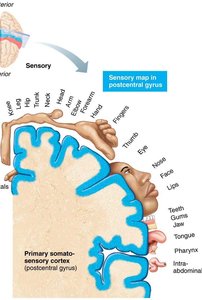
Sensory Areas
Primary somatosensory cortex: Receives sensory information from skin and proprioceptors; capable of spatial discrimination. Somatosensory homunculus represents contralateral sensory input.
Somatosensory association cortex: Integrates sensory input for understanding objects; determines size, texture, and relationship of parts.
Visual Areas
Primary visual cortex: Located in the occipital lobe; receives visual information from retinas. Damage results in functional blindness.
Visual association area: Surrounds primary visual cortex; interprets visual stimuli using past experiences. Damage results in inability to comprehend visual information.

Auditory, Vestibular, Olfactory, Gustatory, and Visceral Sensory Areas
Primary auditory cortex: Interprets information from inner ear as pitch, loudness, and location.
Auditory association area: Stores memories of sounds and permits perception of sound stimulus.
Vestibular cortex: Responsible for conscious awareness of balance.
Primary olfactory cortex: Involved in conscious awareness of odors.
Gustatory cortex: Involved in perception of taste.
Visceral sensory area: Conscious perception of visceral sensations.

Multimodal Association Areas
Multimodal association areas receive inputs from multiple sensory areas and send outputs to multiple areas, allowing integration of information, memory storage, and decision-making.
Anterior association area: Involved with intellect, cognition, recall, and personality.
Posterior association area: Recognizes patterns and faces, localizes us in space, and is involved in language comprehension (Wernicke’s area).
Limbic association area: Provides emotional impact and helps establish memories.
Lateralization and Cerebral Dominance
Lateralization: Division of labor between hemispheres.
Hemispheres communicate via fiber tracts and functional integration.
Cerebral dominance: Refers to the hemisphere dominant for language (90% left-sided dominance).
Left hemisphere: language, math, logic; right hemisphere: visual-spatial skills, intuition, emotion, artistic and musical skills.

Cerebral White Matter
Structure and Function
Cerebral white matter is responsible for communication between cerebral areas and between the cortex and lower CNS.
Consists of myelinated fibers bundled into large tracts.
Classified by direction:
Association fibers: Connect different parts of the same hemisphere.
Commissural fibers: Connect gray matter of two hemispheres.
Projection fibers: Connect hemispheres with lower brain or spinal cord.
Basal Nuclei (Ganglia)
Structure and Function
Basal nuclei are deep gray matter structures involved in motor control and cognition.
Include caudate nucleus, putamen, and globus pallidus.
Associated with subthalamic nuclei and substantia nigra.
Functions: influence muscle movements, regulate movement intensity, filter out incorrect responses, inhibit unnecessary movements, and play a role in cognition and emotion.
Diencephalon
Thalamus
Acts as a relay station for information entering the cortex.
Sorts, edits, and relays ascending input for emotion, visceral function, motor control, memory, and sensory integration.
Hypothalamus
Main visceral control and regulating center vital to homeostasis.
Controls autonomic nervous system, initiates physical responses to emotions, regulates body temperature, hunger, thirst, sleep-wake cycles, and endocrine functions.
Epithalamus
Contains the pineal gland, which secretes melatonin to regulate sleep-wake cycles.
Brain Stem
Structure and Function
The brain stem consists of the midbrain, pons, and medulla oblongata.
Controls automatic behaviors necessary for survival.
Contains fiber tracts connecting higher and lower neural centers.
Midbrain
Cerebral peduncles contain pyramidal motor tracts.
Corpora quadrigemina: superior colliculi (visual reflex), inferior colliculi (auditory relay).
Substantia nigra and red nucleus are involved in motor pathways.
Pons
Contains conduction tracts connecting higher brain centers and spinal cord.
Relays impulses between motor cortex and cerebellum.
Helps maintain normal rhythm of breathing.
Medulla Oblongata
Pyramids formed by pyramidal tracts; decussation occurs here.
Olives relay stretch information from muscles and joints to cerebellum.
Contains autonomic reflex centers for cardiovascular, respiratory, and other functions.
Cerebellum
Structure and Function
The cerebellum processes input from the cortex, brain stem, and sensory receptors to coordinate skeletal muscle movements and maintain balance.
Contains a thin cortex of gray matter and a distinctive treelike pattern of white matter called arbor vitae.
Cerebellar peduncles connect the cerebellum to the brain stem.
All fibers are ipsilateral.
Cerebellar Processing
Receives impulses from the cerebral cortex to initiate voluntary muscle contraction.
Receives signals from proprioceptors, visual, and equilibrium pathways.
Calculates the best way to coordinate muscle contraction.
Sends a "blueprint" of movement to the cerebral motor cortex and brain stem nuclei.
Functional Brain Systems
Limbic System
Responsible for emotional responses and memory formation.
Includes amygdaloid body, cingulate gyrus, hippocampus.
Reticular Formation
Governs brain arousal and consciousness via the reticular activating system (RAS).
Filters out repetitive stimuli; injury can result in coma.
Regulates visceral motor functions.
Higher Mental Functions
Language
Broca’s area: speech production; Wernicke’s area: language comprehension.
Lesions in Broca’s area result in inability to speak; lesions in Wernicke’s area result in nonsensical speech.
Memory
Types: declarative (facts), procedural (skills), motor, emotional.
Short-term memory (STM) and long-term memory (LTM).
Factors affecting transfer: emotional state, rehearsal, association, automatic memory.
Brain Waves and EEG
EEG records electrical activity; waves classified as alpha, beta, theta, delta.
Consciousness and Sleep
Consciousness graded as alertness, drowsiness, stupor, coma.
Sleep includes NREM and REM stages; regulated by hypothalamic nuclei.
Protection of the Brain
Meninges
Three layers: dura mater, arachnoid mater, pia mater.
Functions: protect CNS, enclose venous sinuses, contain CSF, form partitions.
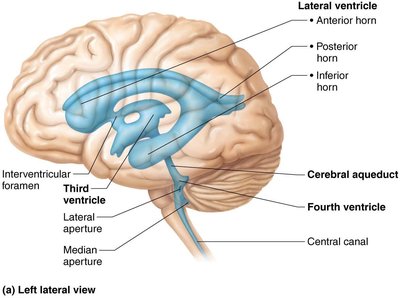
Cerebrospinal Fluid (CSF)
CSF forms a liquid cushion, protects CNS, nourishes brain, and removes waste.
Produced by choroid plexus; flows through ventricles and subarachnoid space.
Blood Brain Barrier
Maintains stable environment; tight junctions prevent passage of harmful substances.
Allows passage of lipid-soluble substances and blood gases; uses specific transport mechanisms for glucose and other important molecules.
Brain Dysfunction
Traumatic Brain Injuries
Includes concussion, contusion, hemorrhage, cerebral edema.
Cerebrovascular Accidents (Strokes)
Ischemia (blood supply deprivation), hemiplegia, TIAs, tPA treatment.
Degenerative Brain Disorders
Alzheimer’s disease: progressive dementia, beta-amyloid plaques, neurofibrillary tangles.
Parkinson’s disease: degeneration of dopamine neurons, tremors.
Huntington’s disease: hereditary, degeneration of basal nuclei and cortex, wild movements.
Spinal Cord: Structure and Function
Gross Anatomy and Protection
Enclosed in vertebral column; begins at foramen magnum, ends at L1/L2.
31 pairs of spinal nerves; major reflex center.
Spinal dura mater, epidural space, CSF in subarachnoid space.
Cross-Sectional Anatomy
Gray matter in core, white matter outside.
Gray matter: dorsal horns (sensory), ventral horns (motor), lateral horns (sympathetic), gray commissure.
Spinal roots: ventral (motor), dorsal (sensory), dorsal root ganglia.
White matter: ascending (sensory), descending (motor), transverse (commissural) fibers; divided into dorsal, lateral, ventral columns.
Spinal Cord Trauma and Disorders
Paresthesias (sensory loss), paralysis (motor loss), flaccid and spastic paralysis, transection (paraplegia, quadriplegia).
Poliomyelitis: destruction of ventral horn motor neurons by poliovirus.
Amyotrophic lateral sclerosis (ALS): destruction of motor neurons and pyramidal tract fibers.
Neuronal Pathways
Ascending Pathways
Conduct sensory information upward through three neurons: first-order, second-order, third-order.
Main pathways: dorsal column–medial lemniscal, spinothalamic, spinocerebellar tracts.
Descending Pathways
Deliver motor impulses from brain to spinal cord; involve upper and lower motor neurons.
Direct (pyramidal) pathways: fast, fine movements.
Indirect pathways: reticulospinal, vestibulospinal, rubrospinal, tectospinal tracts.
Developmental Aspects of CNS
Neural plate forms neural groove and neural tube; neural crest forms dorsal root ganglia.
Alar plate becomes interneurons; basal plate becomes motor neurons.
Clinical Homeostatic Imbalances
Cerebral palsy: neuromuscular disability from brain damage.
Anencephaly: failure of neural fold fusion; fatal.
Spina bifida: incomplete vertebral arch formation; may cause neural problems.
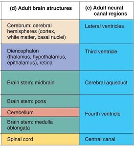
Adult Brain Structure | Adult Neural Canal Region |
|---|---|
Cerebrum: cerebral hemispheres (cortex, white matter, basal nuclei) | Lateral ventricles |
Diencephalon (thalamus, hypothalamus, epithalamus, retina) | Third ventricle |
Brain stem: midbrain | Cerebral aqueduct |
Brain stem: pons | Fourth ventricle |
Cerebellum | Fourth ventricle |
Brain stem: medulla oblongata | Fourth ventricle |
Spinal cord | Central canal |
Additional info: This table summarizes the relationship between adult brain structures and their corresponding neural canal regions.
