Back
BackCentral Nervous System: Structure, Function, and Integration
Study Guide - Smart Notes
 Tailored notes based on your materials, expanded with key definitions, examples, and context.
Central Nervous System (CNS)
Overview of CNS Functions
The central nervous system (CNS) is responsible for integrating sensory information, making decisions, and coordinating motor output. It consists of the brain and spinal cord, which work together to maintain homeostasis and enable complex behaviors.
Sensory Functions: Detection of sensations inside and outside the body.
Integrative Functions: Decision-making processes, primarily within the CNS.
Motor Functions: Stimulation of muscle contractions or gland secretions.
Sensory and motor functions are performed by the peripheral nervous system (PNS), while integrative functions are exclusive to the CNS.
Basic Structure of the Brain and Spinal Cord
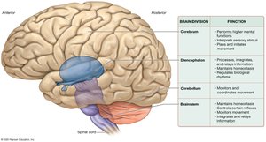
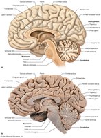
The brain is a soft, whitish-gray organ located in the cranial cavity, divided into four main regions. It contains internal cavities called ventricles, filled with cerebrospinal fluid (CSF), and receives about 20% of the body's blood flow at rest.
Cerebrum: Responsible for higher mental functions, sensation, and movement.
Diencephalon: Processes and relays information, maintains homeostasis, and regulates movement and rhythms.
Cerebellum: Coordinates movement and balance.
Brainstem: Controls basic involuntary processes and connects the brain to the spinal cord.
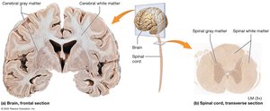
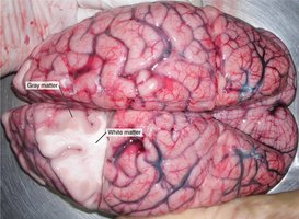
White Matter and Gray Matter
White matter consists of myelinated axons, while gray matter is composed of neuron cell bodies, dendrites, and unmyelinated axons. In the brain, gray matter forms the outer cortex and is scattered in deeper regions, while in the spinal cord, gray matter is internal and white matter is superficial.
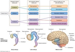
Development of the CNS
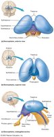
The CNS develops from a hollow neural tube, which forms primary brain vesicles (forebrain, midbrain, hindbrain) that later differentiate into secondary vesicles and mature brain structures.
The Cerebrum
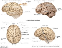
Structure and Lobes
The cerebrum consists of two hemispheres, each divided into lobes: frontal, parietal, temporal, occipital, and insula. These lobes are separated by sulci (shallow grooves) and fissures (deep grooves).
Cerebral Cortex and Functional Areas
The neocortex is the most recent evolutionary part of the cerebral cortex, responsible for conscious processes. It contains primary motor and sensory cortices, as well as association areas for integrating stimuli.
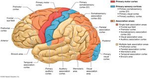
Primary Motor Cortex: Plans and executes movement.
Primary Sensory Cortices: Process sensory input (e.g., somatosensory, visual, auditory).
Association Areas: Integrate multiple types of stimuli.
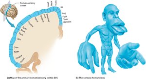
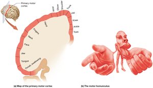
Somatotopy and Motor/Sensory Homunculus
The primary somatosensory and motor cortices are organized somatotopically, meaning specific regions correspond to specific body parts. This is depicted in the sensory and motor homunculus maps.
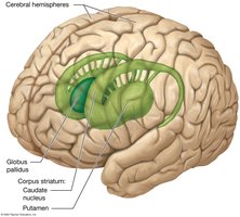
Basal Nuclei
The basal nuclei are clusters of cell bodies within the cerebral hemispheres that help inhibit involuntary movement and initiate voluntary movement. Major components include the caudate nucleus, putamen, and globus pallidus.
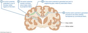
Cerebral White Matter
Commissural tracts: Connect right and left hemispheres (e.g., corpus callosum).
Projection tracts: Connect cortex with other brain regions and spinal cord.
Association tracts: Connect regions within a single hemisphere.
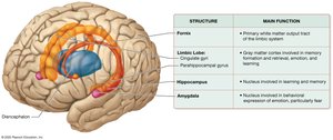
Limbic System
The limbic system, found only in mammals, is involved in memory, learning, emotion, and behavior. Key structures include the limbic lobe, hippocampus, amygdala, and fornix.
The Diencephalon
Major Components
Thalamus: Main relay station for sensory information to the cortex.
Hypothalamus: Regulates homeostasis, autonomic nervous system, and endocrine system; connects to the pituitary gland.
Epithalamus: Contains the pineal gland, which secretes melatonin.
Subthalamus: Works with basal nuclei to control movement.
The Cerebellum
Structure and Function
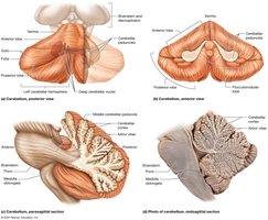
The cerebellum is located posteriorly and inferiorly, with two hemispheres connected by the vermis. It coordinates movement, motor memory, posture, and balance.
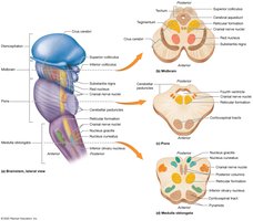
The Brainstem
Major Divisions and Functions
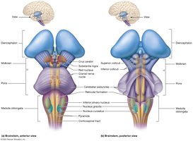
Midbrain: Controls movement, visual and auditory reflexes.
Pons: Regulates breathing, sleep, and arousal.
Medulla Oblongata: Contains centers for cardiovascular, respiratory, and reflex functions.
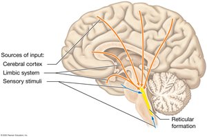
Reticular Formation: Maintains alertness and regulates sleep-wake cycles.
Protection of the Brain
Cranial Meninges
The brain is protected by three layers of meninges: dura mater (outermost), arachnoid mater (middle), and pia mater (innermost). These layers provide structural support and contain spaces filled with CSF.
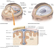
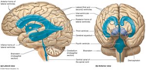
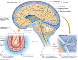
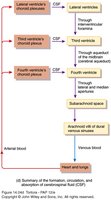
Ventricles and Cerebrospinal Fluid (CSF)
The brain contains four ventricles filled with CSF, which cushions the brain, removes waste, and maintains homeostasis. CSF circulates through the ventricles and subarachnoid space, and is reabsorbed into the blood by arachnoid villi.
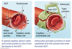
Blood Brain Barrier
The blood brain barrier is formed by endothelial cells with tight junctions, astrocyte foot processes, and a basal lamina. It restricts the passage of large or polar substances, protecting the brain from toxins but also limiting drug delivery.
The Spinal Cord
Structure and Function
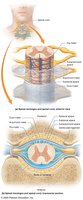
The spinal cord acts as a relay and processing station, transmitting signals between the brain and body and mediating spinal reflexes. It is protected by spinal meninges and surrounded by vertebrae.
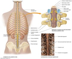
Internal Anatomy
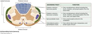
The spinal cord contains a central canal filled with CSF, surrounded by gray matter (butterfly-shaped) and white matter (funiculi). The gray matter contains ventral (motor), dorsal (sensory), and lateral (autonomic) horns.
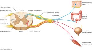
Sensation and Perception
General Somatic Senses
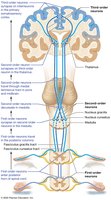
General somatic senses include touch, stretch, joint position, pain, and temperature. Sensory pathways involve first-order (PNS), second-order (spinal cord/brainstem), and third-order (thalamus to cortex) neurons.
Special Senses
Special senses include vision, hearing, taste, smell, and balance. Each sense has dedicated pathways and cortical areas for processing and perception.
Voluntary Movement
Motor Pathways
Voluntary movement is planned and coordinated by the cerebral cortex, basal nuclei, cerebellum, and spinal cord. Upper motor neurons originate in the cortex, while lower motor neurons innervate skeletal muscles.
Learning, Memory, and Cognition
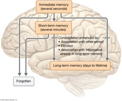
Memory Types
Immediate Memory: Lasts seconds; used in conversation and daily tasks.
Short-Term Memory: Lasts minutes; allows manipulation of information.
Long-Term Memory: Lasts days to a lifetime; consolidation is required for storage.
Cognition and Language
Cognition involves higher-order functions such as awareness, reasoning, and personality, primarily managed by the prefrontal cortex. Language is processed in Broca's and Wernicke's areas, mainly in the left hemisphere.
Homeostasis and Sleep
Homeostatic Regulation
The hypothalamus is the primary regulator of homeostasis, controlling body temperature, hunger, thirst, and the autonomic nervous system. The reticular formation regulates sleep-wake cycles and consciousness.
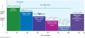
Sleep and Brain Waves
Sleep consists of non-REM and REM stages, each with characteristic brain wave patterns. Sleep is essential for brain function, memory consolidation, and metabolic waste clearance.