 Back
BackCentral Nervous System: Structure, Function, and Development
Study Guide - Smart Notes
 Tailored notes based on your materials, expanded with key definitions, examples, and context.
The Central Nervous System
Emergent Properties of Neural Networks
The central nervous system (CNS) exhibits complex behaviors and functions that arise from the interactions of neural networks. These emergent properties include plasticity, affective and cognitive behaviors, learning, and memory.
Plasticity: The ability of the brain to reorganize neural pathways based on sensory input and experience.
Affective behaviors: Related to feelings and emotions.
Cognitive behaviors: Related to thinking and reasoning.
Learning: The acquisition of knowledge or skills through experience.
Memory: The retention and recall of information.
Evolution of Nervous Systems
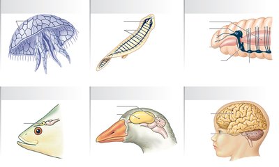
The nervous system has evolved from simple structures in unicellular organisms to complex brains in vertebrates. This evolution is closely linked to behavioral complexity.
Unicellular organisms: Use membrane potentials for activity coordination.
Cnidaria: Possess a nerve net.
Flatworms: Have primitive brains and nerve cords.
Annelids: Feature simple brains and ganglia along nerve cords.
Vertebrates: Exhibit dramatic changes in the forebrain, especially the cerebrum.
Anatomy and Development of the CNS
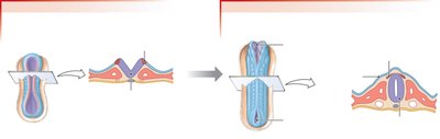
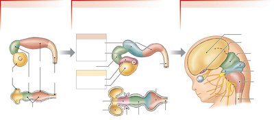
The CNS develops from a hollow neural tube, which differentiates into specialized regions during embryogenesis. The process involves the formation of the neural plate, neural tube, and subsequent division into forebrain, midbrain, and hindbrain.
Neural plate: Group of cells that form the CNS.
Neural tube: Fused plate forms the tube by day 23.
Week 4: Differentiation into forebrain, midbrain, hindbrain.
Week 6: Seven divisions present; formation of ventricles.
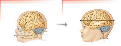
Week 11: Cerebrum enlarges and covers other regions.
Gross Anatomy of the CNS
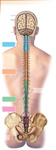
The CNS consists of the brain and spinal cord, protected by bone and connective tissue. The brain is encased in the cranium, and the spinal cord runs through the vertebral column.
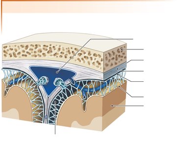
Meninges: Three layers (dura mater, arachnoid membrane, pia mater) stabilize and protect neural tissue.
Gray matter: Unmyelinated cell bodies, dendrites, axon terminals; organized into nuclei.
White matter: Myelinated axons; organized into tracts.
Cerebrospinal Fluid (CSF) and Ventricles
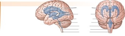
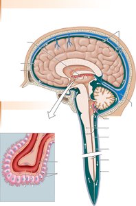
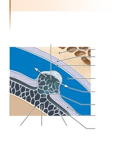
CSF is a salty solution produced by the choroid plexus in the ventricles. It surrounds the brain, providing physical and chemical protection, and is contained within the subarachnoid space.
Production: Choroid plexus selectively moves materials from plasma to ventricles.
Flow: CSF flows from ventricles to subarachnoid space and returns to plasma via villi.
Functions: Cushions CNS, regulates ion concentrations, removes toxins.
The Blood-Brain Barrier (BBB)
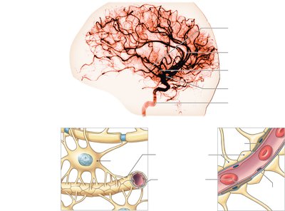
The BBB is a highly selective barrier formed by tight junctions between endothelial cells, promoted by astrocyte foot processes. It protects the brain from toxins and pathogens, allowing only certain substances to cross.
Permeability: Small lipid-soluble molecules and gases cross easily; water-soluble compounds are restricted.
Metabolic requirements: Neurons require constant oxygen and glucose supply.
The Spinal Cord
Structure and Function
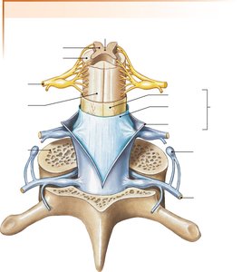
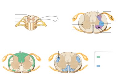
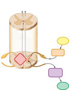
The spinal cord is segmented and associated with spinal nerves. It integrates sensory and motor information and mediates reflexes.
Dorsal root: Carries sensory (afferent) information to CNS.
Ventral root: Carries motor (efferent) information to muscles and glands.
Gray matter: Contains sensory and motor nuclei.
White matter: Divided into columns of tracts (ascending, descending, propriospinal).
Spinal reflexes: Integrated in the spinal cord without brain input.
The Brain
Brain Stem
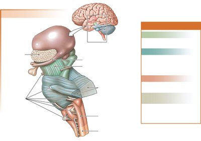
The brain stem is the oldest part of the brain and is responsible for basic life functions. It includes the medulla oblongata, pons, and midbrain.
Medulla oblongata: Controls involuntary functions (blood pressure, breathing, swallowing, vomiting).
Pons: Relay station, coordinates breathing.
Midbrain: Controls eye movement, relays signals for hearing and seeing reflexes.
Reticular formation: Controls wakefulness, sleep, muscle tone, pain modulation.
Cerebellum
The cerebellum is the second largest brain structure, responsible for coordinating movement, equilibrium, and balance. It processes sensory information and motor input from the cerebrum.
Muscle memory: The cerebellum is involved in procedural learning.
Alcohol: Inhibits cerebellar function.
Diencephalon
The diencephalon contains centers for homeostasis and behavioral drives. It includes the thalamus, hypothalamus, pituitary gland, and pineal gland.
Thalamus: Relay station and integrating center for sensory and motor information.
Hypothalamus: Controls homeostasis, behavioral drives, autonomic and endocrine functions.
Pituitary gland: Secretes hormones.
Pineal gland: Secretes melatonin, regulates circadian rhythms.


Function | Structure |
|---|---|
Homeostasis | Hypothalamus |
Relay station | Thalamus |
Hormone secretion | Pituitary gland |
Circadian rhythm | Pineal gland |
Behavioral drives | Hypothalamus |
Cerebrum
The cerebrum is the site of higher brain functions, including cognition, affect, behavior, and personality. It consists of two hemispheres connected by the corpus callosum.
Gray matter: Includes cerebral cortex, basal ganglia (movement control), limbic system (emotion and memory).
White matter: Bundles of fibers connecting brain regions.
Cerebral lateralization: Functional asymmetry between hemispheres (left: math/language; right: visual/spatial).
Brain Function
Functional Areas of the Cerebral Cortex
The cerebral cortex is organized into sensory, motor, and association areas, each responsible for specific functions.
Sensory areas: Translate sensory input into perception.
Motor areas: Direct skeletal muscle movement.
Association areas: Integrate information and direct voluntary behaviors.
Diffuse Modulatory Systems
Neurons in the reticular formation project to large areas of the brain, modulating sensory and cognitive processes. These systems use neurotransmitters such as norepinephrine, serotonin, dopamine, and acetylcholine.
Sleep and Circadian Rhythms
Sleep is regulated by the brain and consists of non-REM and REM phases. The suprachiasmatic nucleus (SCN) of the hypothalamus acts as the primary clock for circadian rhythms.
REM sleep: Characterized by dreaming and muscle paralysis.
Sleep disorders: Include insomnia, sleep apnea, and somnambulism.
Emotion, Motivation, and Memory
The limbic system is the center of emotion and motivation. Memory is classified as short-term, working, and long-term, with further division into reflexive (implicit) and declarative (explicit) memory.
Type | Recall | Acquisition | Storage |
|---|---|---|---|
Reflexive (Implicit) | Automatic | Repetition | Cerebellum |
Declarative (Explicit) | Conscious | Higher-level thinking | Cerebral cortex |
Language and Higher Cognitive Functions
Language processing involves Wernicke's area (comprehension) and Broca's area (expression). Damage to these areas results in aphasia.
Wernicke's area: Understanding language.
Broca's area: Producing speech.
Personality
Personality is shaped by both genetic and environmental factors. Disorders such as schizophrenia have both bases.
Cranial Nerves
The brain stem gives rise to 12 cranial nerves, each with specific sensory, motor, or mixed functions.
Number | Name | Type | Primary Function |
|---|---|---|---|
I | Olfactory | Sensory | Smell |
II | Optic | Sensory | Vision |
III | Oculomotor | Motor | Eye movement, pupil constriction |
IV | Trochlear | Motor | Eye movement |
V | Trigeminal | Mixed | Sensory from face, motor for chewing |
VI | Abducens | Motor | Eye movement |
VII | Facial | Mixed | Taste, facial expression |
VIII | Vestibulocochlear | Sensory | Hearing, equilibrium |
IX | Glossopharyngeal | Mixed | Swallowing, salivary secretion |
X | Vagus | Mixed | Internal organs, muscles, glands |
XI | Spinal accessory | Motor | Neck, shoulder muscles |
XII | Hypoglossal | Motor | Tongue muscles |
Summary
The central nervous system is a highly organized structure responsible for integrating sensory information, coordinating motor output, and supporting higher cognitive functions. Its development, anatomy, and function are foundational to understanding human physiology.
