 Back
Central Nervous System: The Brain – Structure, Function, and Protection
Study Guide - Smart Notes
 Tailored notes based on your materials, expanded with key definitions, examples, and context.
CNS: The Brain
Overview of the Central Nervous System
The central nervous system (CNS) consists of the brain and spinal cord, serving as the main control center for the body. The brain is responsible for processing sensory information, coordinating movement, and regulating homeostasis.
Structure: The brain is divided into four main regions: cerebral hemispheres, diencephalon, brain stem, and cerebellum.
Function: Each region has specialized roles in sensory processing, motor control, cognition, and autonomic regulation.
Homeostasis: The CNS maintains internal balance through neural and endocrine mechanisms.
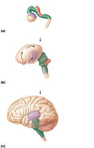
Brain Development and Regions
The brain develops from embryonic regions that fold and grow to form the adult structure.
Telencephalon: Forms the cerebral hemispheres.
Diencephalon: Forms the thalamus, hypothalamus, and epithalamus.
Mesencephalon: Forms the midbrain.
Metencephalon: Forms the pons and cerebellum.
Myelencephalon: Forms the medulla oblongata.
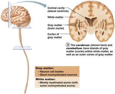
Basic Pattern of the CNS: Gray and White Matter
The CNS is organized into gray and white matter.
Gray Matter: Contains neuron cell bodies and short, nonmyelinated neurons.
White Matter: Composed of myelinated axons, facilitating communication between regions.
Cortex: The outer layer of gray matter in the cerebral hemispheres and cerebellum.
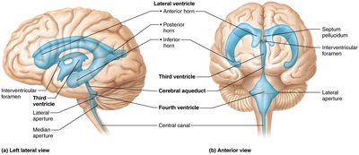
Ventricles of the Brain
The brain contains fluid-filled chambers called ventricles, which are continuous with the central canal of the spinal cord.
Lateral Ventricles: Paired, C-shaped chambers in the cerebral hemispheres.
Third Ventricle: Located in the diencephalon.
Fourth Ventricle: Located dorsal to the pons and medulla in the hindbrain.
Function: Ventricles are filled with cerebrospinal fluid (CSF) and lined by ependymal cells.
Cerebral Hemispheres
Structure and Lobes
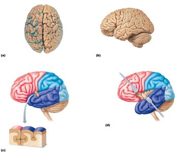
The cerebral hemispheres form the superior part of the brain and are divided into lobes by sulci and fissures.
Lobes: Frontal, parietal, temporal, occipital, and insula.
Gyri: Ridges on the surface.
Sulci: Shallow grooves.
Fissures: Deep grooves separating major regions.
Functional Areas of the Cerebral Cortex
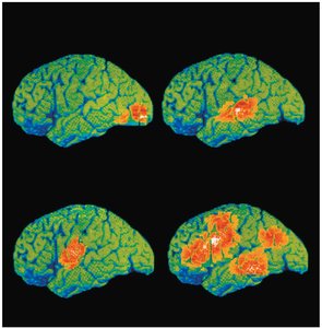
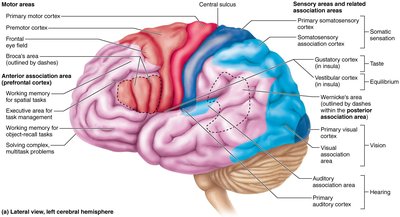
The cerebral cortex is the site of conscious mind, enabling sensation, communication, memory, understanding, and voluntary movements.
Motor Areas: Control voluntary movement (e.g., primary motor cortex, premotor cortex, Broca’s area, frontal eye field).
Sensory Areas: Provide conscious awareness of sensation (e.g., primary somatosensory cortex, visual, auditory, gustatory, olfactory, vestibular areas).
Association Areas: Integrate diverse information for higher functions (e.g., prefrontal cortex, posterior association area, limbic association area).
Motor and Sensory Maps
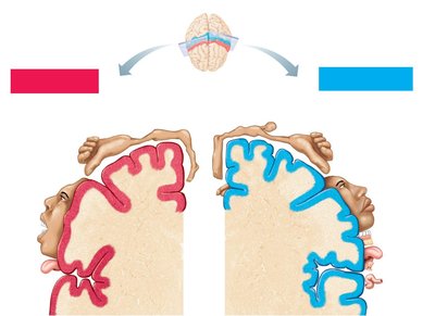
The primary motor and somatosensory cortices are organized as body maps, representing different regions of the body.
Motor Map: Located in the precentral gyrus.
Sensory Map: Located in the postcentral gyrus.
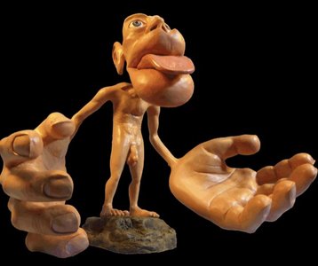
Homunculus: Visual representation of the relative space devoted to each body part.
Lateralisation of Cortical Function
Each hemisphere of the brain has specialized functions.
Left Hemisphere: Dominant for language, math, and logic (90% of humans).
Right Hemisphere: Controls visual-spatial skills, emotion, artistic and musical abilities.
Cerebral Dominance: Refers to the hemisphere dominant for language.
Cerebral White Matter
White matter in the cerebral hemispheres is responsible for communication between regions.
Commissures: Connect corresponding gray areas of the two hemispheres (e.g., corpus callosum).
Association Fibers: Connect different parts of the same hemisphere.
Projection Fibers: Connect the cortex with lower brain or spinal cord centers.
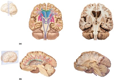
Basal Nuclei
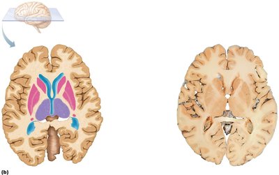
Basal nuclei are masses of gray matter deep within the white matter of the cerebrum.
Components: Caudate nucleus, putamen, globus pallidus.
Functions: Influence muscular activity, regulate attention and cognition, inhibit unnecessary movements.
Clinical Relevance: Disorders such as Parkinson’s and Huntington’s disease are associated with basal nuclei dysfunction.
Diencephalon
Structure and Components
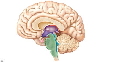
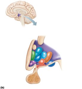
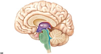
The diencephalon is the central core of the forebrain, consisting of three paired gray matter structures: thalamus, hypothalamus, and epithalamus.
Thalamus: Relay station for sensory and motor information.
Hypothalamus: Main visceral control center, regulates homeostasis and links nervous and endocrine systems.
Epithalamus: Contains the pineal gland, which secretes melatonin for sleep regulation.
Thalamus
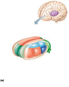
The thalamus contains multiple nuclei that sort, edit, and relay information to the cerebral cortex.
Functions: Mediates sensation, motor activities, cortical arousal, learning, and memory.
Nuclei: Anterior, ventral, dorsal, posterior, medial, lateral, and reticular nuclei.
Hypothalamus
The hypothalamus is vital for homeostasis, controlling autonomic functions, emotional responses, body temperature, hunger, thirst, sleep-wake cycles, and endocrine activity.
Mammillary Bodies: Involved in olfactory relay.
Infundibulum: Connects hypothalamus to the pituitary gland.
Hormones: Produces ADH and oxytocin.
Epithalamus
The epithalamus forms the roof of the third ventricle and contains the pineal gland and choroid plexus.
Pineal Gland: Secretes melatonin, regulating sleep-wake cycles and mood.
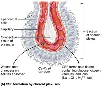
Choroid Plexus: Produces cerebrospinal fluid (CSF).
Brain Stem
Structure and Functions
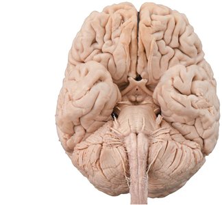
The brain stem consists of the midbrain, pons, and medulla oblongata. It controls automatic behaviors necessary for survival and serves as a pathway for neural tracts.
Midbrain: Contains nuclei for cranial nerves III and IV, visual and auditory reflex centers, substantia nigra, and red nucleus.
Pons: Connects higher brain centers and the spinal cord, relays impulses between motor cortex and cerebellum, origin of cranial nerves V, VI, VII.
Medulla Oblongata: Autonomic reflex center, cardiovascular and respiratory control, origin of cranial nerves X, XI, XII.
Cerebellum
Structure and Function
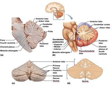
The cerebellum is located dorsal to the pons and medulla, under the occipital lobes. It coordinates voluntary movements, balance, and posture.
Lobes: Anterior, posterior, and flocculonodular.
Arbor Vitae: Treelike pattern of white matter.
Function: Provides precise timing and patterns of skeletal muscle contraction, plays a role in language and problem solving.
Functional Brain Systems
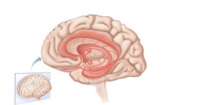
Limbic System
The limbic system is involved in emotion, memory, and motivation.
Components: Amygdala, cingulate gyrus, hippocampus, hypothalamus, anterior thalamic nucleus.
Functions: Emotional responses, memory formation, psychosomatic illnesses.
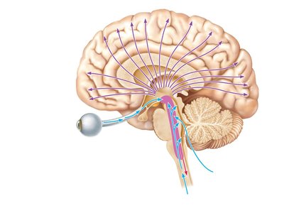
Reticular Formation
The reticular formation is a network of neurons in the brain stem that regulates arousal and consciousness.
Reticular Activating System (RAS): Keeps the cerebral cortex alert, filters sensory input.
Clinical Relevance: Injury can result in coma.
Higher Mental Functions
Language, Memory, Consciousness, and Sleep
Higher mental functions are processed in specialized cortical areas.
Language: Involves Broca’s and Wernicke’s areas.
Memory: Storage and retrieval, short-term and long-term memory, hippocampus involvement.
Consciousness: Awareness of self and environment.
Sleep: Regulated by the hypothalamus and reticular formation.
Protection of the Brain
Skull, Meninges, Cerebrospinal Fluid, and Blood-Brain Barrier
The brain is protected by multiple layers and mechanisms.
Skull: Provides rigid protection.
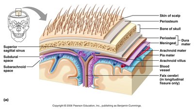
Meninges: Three connective tissue membranes (dura mater, arachnoid mater, pia mater) cover and protect the CNS, enclose blood vessels, and contain CSF.
Cerebrospinal Fluid (CSF): Cushions the brain, nourishes it, and removes waste.
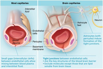
Blood-Brain Barrier: Selective barrier that protects the brain from harmful substances while allowing essential nutrients to pass.
Summary Table: Major Brain Regions and Functions
Region | Main Function |
|---|---|
Cerebral Hemispheres | Conscious thought, voluntary movement, sensory processing |
Diencephalon | Relay sensory/motor info, homeostasis, endocrine regulation |
Brain Stem | Autonomic functions, cranial nerve origin, neural pathways |
Cerebellum | Coordination, balance, motor learning |
Key Equations and Concepts
Neural Transmission: (Ohm's Law for neural conduction; V = voltage, I = current, R = resistance)
CSF Formation:
Additional info:
Memory formation and retrieval involve complex biochemical and structural changes in neurons.
The blood-brain barrier is absent in certain regions (e.g., hypothalamus) to allow monitoring of blood chemistry.
