 Back
Central Nervous System: The Spinal Cord – Structure, Function, and Nerve Plexuses
Study Guide - Smart Notes
 Tailored notes based on your materials, expanded with key definitions, examples, and context.
Central Nervous System: The Spinal Cord
Introduction
The spinal cord is a major component of the central nervous system (CNS), responsible for transmitting and processing information between the brain and the rest of the body. This section covers the anatomical features, protective coverings, internal structure, and nerve plexuses associated with the spinal cord.
Longitudinal Anatomy of the Spinal Cord
Regions and Structure
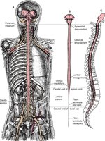
The adult spinal cord is approximately eighteen inches long, extending from the foramen magnum to the first lumbar vertebra. It is divided into regions corresponding to the vertebral column: cervical, thoracic, lumbar, and sacral. Two enlargements, cervical and lumbar, accommodate increased neural structures for the limbs.
Cervical enlargement: C3–T2, nerves to/from arms
Lumbar enlargement: T7–T11, nerves to/from legs
Cauda equina: Bundle of nerves resembling a horse’s tail, extending beyond the spinal cord
Conus medullaris: Terminal end of the spinal cord at L1
Terminal filum: Thin strand anchoring the spinal cord to the coccyx
Spinal nerves: 31 pairs, named by region (C1–C8, T1–T12, L1–L5, S1–S5, 1 coccygeal)
The Meninges
Protective Coverings
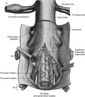
The spinal cord and brain are protected by three layers of meninges:
Dura mater: Outermost, tough and durable, anchored to vertebral cavity
Arachnoid mater: Middle, thin, web-like, connects dura to pia mater
Pia mater: Innermost, thin, rich in blood vessels, directly contacts CNS tissue
The subarachnoid space between arachnoid and pia mater contains cerebrospinal fluid (CSF), which cushions the CNS. CSF can be sampled via lumbar puncture for diagnostic purposes, such as detecting meningitis.
Cross-sectional Anatomy of the Spinal Cord
Gray and White Matter
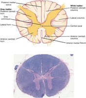
Each spinal cord segment is associated with spinal nerves forming two nerve roots: posterior (dorsal, sensory) and anterior (ventral, motor). The cell bodies of sensory neurons are grouped in the posterior root ganglion.
Gray matter: Butterfly-shaped, divided into horns (posterior, anterior, lateral)
Posterior horn: Receives sensory input
Anterior horn: Sends motor output
Lateral horn: Present in thoracic, upper lumbar, and sacral regions; part of autonomic nervous system
Gray commissure: Connects sides of gray matter, contains central canal
White matter: Organized into columns (posterior, anterior, lateral)
Ascending tracts: Carry sensory information to the brain
Descending tracts: Carry motor commands from the brain
Nerve Plexuses
Organization and Function
Spinal nerves reorganize into nerve plexuses to innervate the periphery. Four major plexuses are found along the vertebral column:
Cervical plexus: C1–C5, branches to neck, head, and phrenic nerve (diaphragm)
Brachial plexus: C5–T1, nerves to arms (radial, median, ulnar, musculocutaneous, axillary)
Lumbar plexus: L1–L4, nerves to pelvic region and anterior leg (femoral, obturator)
Sacral plexus: L4–S4, nerves to posterior leg (sciatic, tibial, fibular)
Thoracic nerves (T2–T11) do not form plexuses but give rise to intercostal nerves for the muscles between ribs.
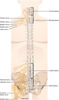
Table: Nerve Plexuses and Associated Nerves
Name | Associated Spinal Nerves | Major Associated Peripheral Nerves |
|---|---|---|
Cervical | C1–C5 | Phrenic |
Brachial | C5–T1 | Radial, median, ulnar, musculocutaneous, axillary |
Lumbar | L1–L4 | Femoral, obturator |
Sacral | L4–S4 | Sciatic |
Key Terms and Concepts
Spinal cord: CNS structure transmitting information between brain and body
Meninges: Protective layers (dura, arachnoid, pia mater)
Cauda equina: Bundle of nerves below spinal cord
Gray matter: Neuronal cell bodies, divided into horns
White matter: Myelinated axons, organized into columns
Nerve plexus: Network of nerves reorganizing axons for peripheral innervation
Example: Sensory and Motor Pathways
For example, the radial nerve contains both sensory fibers (entering via posterior root) and motor fibers (exiting via anterior root) to the arm. The pathway for a sensory signal to excite a muscle via motor nerves involves:
Sensory nerve innervating the muscle
Posterior root
Posterior root ganglion
Posterior horn
Ascending tracts of white matter
Brain
Descending tracts of white matter
Anterior horn
Anterior root
Motor nerve innervating the muscle
Summary
The spinal cord is a complex structure essential for sensory and motor signaling, protected by meninges and organized into regions and nerve plexuses. Understanding its anatomy and function is fundamental for studies in anatomy and physiology.
