 Back
Chapter 1: An Introduction to Anatomy and Physiology – Structured Study Notes
Study Guide - Smart Notes
 Tailored notes based on your materials, expanded with key definitions, examples, and context.
Chapter 1: An Introduction to Anatomy and Physiology
1-2 Anatomy and Physiology
Anatomy and physiology are foundational sciences for understanding the structure and function of the human body. They are closely integrated, as the form of a structure is directly related to its function.
Anatomy: The study of internal and external body structures and their physical relationships among other body parts.
Physiology: The study of how living organisms perform their vital functions.
Principle of Complementarity: All specific functions are performed by specific structures; the form of a structure relates to its function.
Types of Anatomy
Gross (Macroscopic) Anatomy: Examines large structures visible with the naked eye.
Regional Anatomy: Focuses on specific body areas.
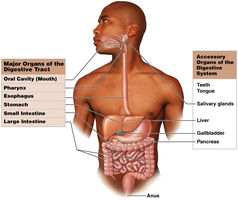
Systemic Anatomy: Studies organ systems.
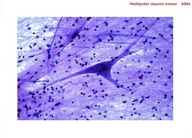
Microscopic Anatomy: Examines structures only visible with magnification, such as cells and molecules.
Cytology: Study of cell structure.
Histology: Study of tissue structure.
Types of Physiology
Cell Physiology: Function of cells and their chemical processes.
Organ Physiology: Function of specific organs.
Systemic Physiology: Function of organ systems.
Pathological Physiology: Effects of diseases on organs or systems.
Clinical Application
Signs: Objective disease indications (e.g., fever).
Symptoms: Subjective disease indications (e.g., tiredness).
Diagnosis: Uses scientific method: observation, hypothesis, experimentation.
1-3 Levels of Organization
The human body is organized into six major levels, from simplest to most complex:
Chemical Level: Atoms and molecules.
Cellular Level: Cells, the smallest living units.
Tissue Level: Groups of cells working together. Four types: epithelial, connective, muscle, neural.
Organ Level: Two or more tissues working together (e.g., heart).
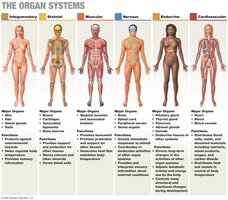
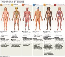
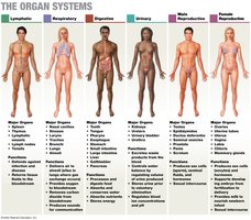
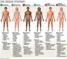
Organ System Level: Groups of organs interacting for a particular function (e.g., cardiovascular system).
Organism Level: An individual life form.
1-5 Anatomical Terminology
Anatomical terminology provides standardized language for describing body regions, sections, and positions.
Anatomical Position: Standard reference position: hands at sides, palms forward, feet together.
Anterior View: Body from the front.
Posterior View: Body from the back.
Supine: Lying face up.
Prone: Lying face down.
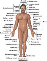
Anatomical Regions
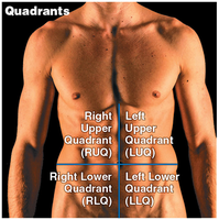
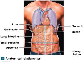
Abdominopelvic Quadrants: Four quadrants used clinically to locate pain or abnormalities: RUQ, LUQ, RLQ, LLQ.
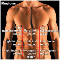
Abdominopelvic Regions: Nine regions used for anatomical studies: epigastric, right/left hypochondriac, umbilical, right/left lumbar, hypogastric, right/left inguinal.
Directional References
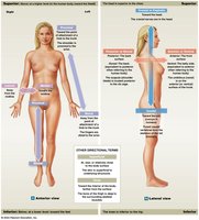
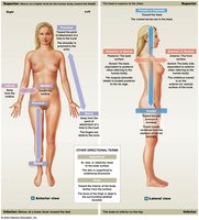
Superior: Toward the head or upper part.
Inferior: Toward the lower part or away from head.
Anterior (Ventral): Nearer to the front.
Posterior (Dorsal): Nearer to the back.
Medial: Nearer to the midline.
Lateral: Toward the side, farther from midline.
Ipsilateral: Same side of the body.
Contralateral: Opposite sides of the body.
Proximal: Nearer to the point of attachment.
Distal: Farther from the point of attachment.
Superficial: Toward or on the surface.
Deep: Away from the surface.
Sectional Anatomy and Medical Imaging
Sectional anatomy involves slicing through a three-dimensional object to visualize internal organization, important for interpreting medical scans.
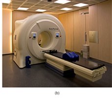
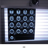
MRI: Noninvasive imaging using magnetic fields and radio waves; does not expose patients to radiation.
fMRI: Detects blood flow concentration to study brain activity.
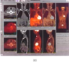
PET: Uses radioactive substances to diagnose conditions; relatively safe.
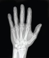
X-ray: High-energy electromagnetic radiation; penetrates solids and ionizes gases.
CT: Analyzes cross-sectional X-rays; exposes patients to higher radiation than X-rays.
Sectional Planes
Frontal (Coronal) Plane: Vertical plane dividing body into anterior and posterior portions.
Sagittal Plane: Vertical plane dividing body into left and right portions. Midsagittal: Middle; Parasagittal: Offset from middle.
Transverse (Horizontal) Plane: Horizontal plane dividing body into superior and inferior portions.
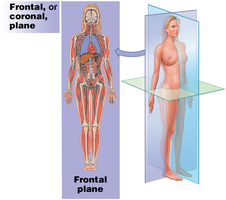
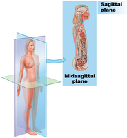
1-6 Body Cavities
Body cavities are closed, fluid-filled spaces lined by serous membranes, containing vital organs (viscera) of the trunk. They protect organs and allow changes in size and shape.
Serous Membrane (Serosa): Lines cavities and covers organs; consists of parietal (lines cavity) and visceral (covers organ) layers; serous fluid reduces friction.
Thoracic Cavity: Divided from abdominopelvic cavity by diaphragm; contains pleural cavities (lungs), pericardial cavity (heart), and mediastinum (connective tissue stabilizing organs).
Abdominopelvic Cavity: Extends from diaphragm to pelvis; contains abdominal cavity, pelvic cavity, and peritoneal cavity (lined with peritoneum).
Retroperitoneal Space: Area between parietal peritoneum and back of body wall; contains pancreas and kidneys.
Pelvic Cavity: Contains reproductive organs, rectum, and urinary bladder.
1-7 Homeostasis
Homeostasis is the continuous physiological process that maintains a stable internal environment. Systems respond to changes to keep variables within normal ranges (e.g., temperature, blood pressure).
Receptor: Sensor detecting stimulus or change.
Control Center: Receives and processes information, sends commands.
Effector: Cell or organ carrying out commands.
Regulatory mechanisms limit fluctuations to keep conditions close to a set point.
1-8 Negative and Positive Feedback
Feedback mechanisms regulate homeostasis:
Negative Feedback: Opposes variation from normal; effector response negates original stimulus; maintains variables within normal range.
Positive Feedback: Enhances variation from normal; response amplifies original change; used for rapid completion of potentially dangerous processes (e.g., blood clotting).
Table: Levels of Organization and Major Organ Systems
Level | Description |
|---|---|
Chemical | Atoms and molecules |
Cellular | Cells, smallest living units |
Tissue | Groups of cells; four types: epithelial, connective, muscle, neural |
Organ | Two or more tissues working together |
Organ System | Groups of organs for a function |
Organism | Individual life form |
Organ System | Major Organs | Functions |
|---|---|---|
Integumentary | Skin, hair, nails | Protection, temperature regulation |
Skeletal | Bones, cartilage | Support, movement, protection |
Muscular | Muscles | Movement, heat production |
Nervous | Brain, spinal cord, nerves | Control, coordination |
Endocrine | Glands | Hormone production, regulation |
Cardiovascular | Heart, blood vessels | Transport, regulation |
Lymphatic | Lymph nodes, vessels | Immunity, fluid balance |
Respiratory | Lungs, airways | Gas exchange |
Digestive | Stomach, intestines | Digestion, absorption |
Urinary | Kidneys, bladder | Excretion, regulation |
Reproductive | Ovaries, testes | Reproduction |
Example: The cardiovascular system includes the heart and blood vessels, which transport blood throughout the body, delivering oxygen and nutrients and removing waste products.
Additional info: Academic context was added to clarify the scientific method, regulatory mechanisms, and the principle of complementarity.
