 Back
BackChapter 1: Introduction to Anatomy and Physiology – Structured Study Notes
Study Guide - Smart Notes
 Tailored notes based on your materials, expanded with key definitions, examples, and context.
Anatomy and Physiology: An Introduction
Definition and Scope
Anatomy is the study of internal and external structures and the physical relationships between body parts. Physiology is the study of the function of those structures. The form of anatomical structures is closely tied to the functions they perform. This course emphasizes anatomy, with additional study of physiology.
Anatomy: Structure of the body
Physiology: Function of body structures
Form and Function: Structure determines function
Subdivisions of Anatomy
Macroscopic (Gross) Anatomy
Macroscopic anatomy involves features visible to the unaided eye and is divided into several approaches:
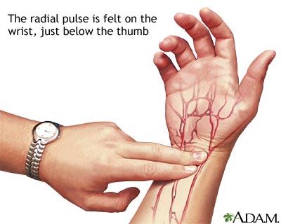
Surface Anatomy: Study of external landmarks that relate to deeper structures. Example: Using wrist landmarks to palpate the radial pulse.
Regional Anatomy: Considers all superficial and internal features in a specific region (e.g., head and neck, thorax).
Systemic Anatomy: Studies the structure of major organ systems that work together to accomplish specific functions (e.g., digestive system, cardiovascular system).
Microscopic Anatomy
Microscopic anatomy examines structures that require magnification:
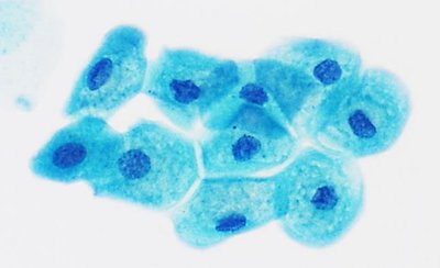
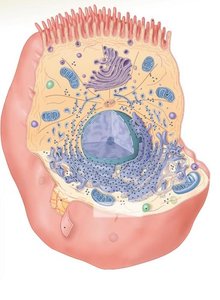
Cytology: Study of individual cells and their internal structures.
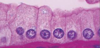
Histology: Study of tissues and how cells are organized to form organs.
Other Anatomical Divisions
Developmental Anatomy: Study of structural changes from conception to death. Embryology focuses on changes from conception to birth.
Pathological Anatomy: Study of how disease affects structures, at both macroscopic and microscopic levels.
Radiographic Anatomy: Study of structures using medical imaging techniques (e.g., X-rays, MRI, ultrasound).
Levels of Organization in the Human Body
Hierarchical Structure
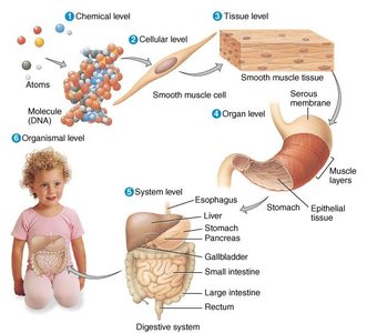
The human body is organized into a hierarchy of increasing complexity:
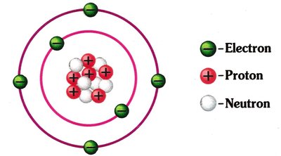
Chemical Level: Atoms and molecules form the basis of all matter.
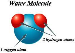
Molecular Level: Atoms bond to form molecules, such as water (H2O).
Cellular Level: Molecules combine to form organelles, which make up cells—the smallest living units.
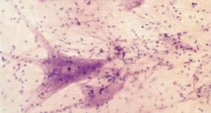
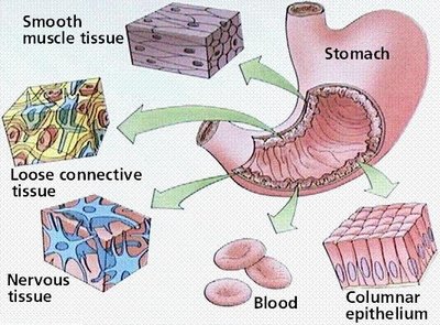
Tissue Level: Similar cells work together to perform specific functions, forming tissues (epithelial, connective, muscle, nervous).

Organ Level: Different tissues combine to form organs, each with specialized functions (e.g., stomach).
Organ System Level: Organs work together in organ systems (e.g., digestive, endocrine).
Organism Level: All organ systems function together to maintain the life of the organism.
Overview of Human Organ Systems
The human body contains 11 major organ systems, each with specific functions:
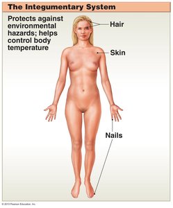
Integumentary System: Protection, temperature regulation
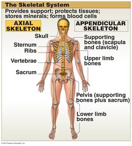
Skeletal System: Support, protection, blood cell formation
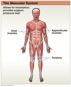
Muscular System: Movement, heat production
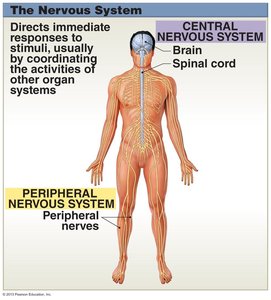
Nervous System: Coordination, response to stimuli
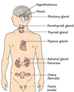
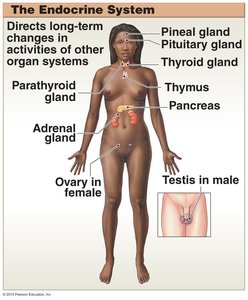
Endocrine System: Hormonal regulation
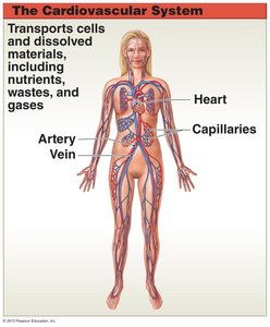
Cardiovascular System: Transport of nutrients, gases, wastes
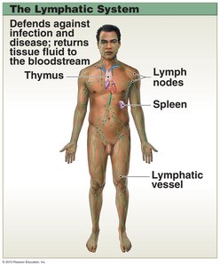
Lymphatic System: Defense, fluid balance
Respiratory System: Gas exchange
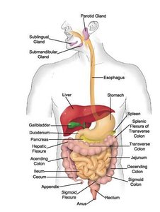
Digestive System: Breakdown and absorption of nutrients
Urinary System: Waste elimination, water balance
Reproductive System: Production of offspring
Homeostasis
Definition and Importance
Homeostasis is the maintenance of a stable internal environment suitable for cellular activities. Disruptions can be caused by external (e.g., temperature, toxins) or internal (e.g., blood pressure, glucose levels) stimuli. Loss of homeostasis can result in illness or death.
Body systems coordinate to maintain homeostasis through homeostatic mechanisms.
Components of Homeostatic Control
Receptor: Senses changes (stimuli) in the environment.
Control Center: Receives information and determines the response (often the CNS or endocrine organs).
Effector: Carries out the response to restore balance (muscles or glands).
Feedback Mechanisms
Negative Feedback: The response opposes the original stimulus, maintaining balance (e.g., body temperature regulation).
Positive Feedback: The response amplifies the original stimulus, used for processes that must be completed quickly (e.g., blood clotting, childbirth).
Anatomical Terminology
Standard Reference Position
The anatomical position is the standard reference for anatomical descriptions: standing erect, feet parallel and forward, arms at sides, palms forward, and thumbs away from the body.
Directional Terms
Directional terms describe the location of one body part relative to another (e.g., superior/inferior, anterior/posterior, medial/lateral, proximal/distal, superficial/deep).
Anatomical Planes
Coronal (Frontal) Plane: Divides the body into anterior and posterior portions.
Sagittal Plane: Divides the body into right and left portions (midsagittal = equal, parasagittal = unequal).
Transverse (Horizontal) Plane: Divides the body into superior and inferior portions.
Body Cavities
Dorsal Cavity: Cranial (brain) and vertebral (spinal cord) cavities.
Ventral Cavity: Thoracic (lungs, heart) and abdominopelvic (digestive, urinary, reproductive organs) cavities.
Serous Membranes
Serous membranes line body cavities and reduce friction. They have two layers: parietal (lines cavity wall) and visceral (covers organ).
Three main serous membranes: Pericardium (heart), Pleura (lungs), Peritoneum (abdominal organs).
Abdominopelvic Quadrants and Regions
The abdominopelvic cavity is divided into quadrants and nine regions for clinical reference and localization of pain or pathology.
Additional info: This summary covers all major introductory concepts in anatomy and physiology, including definitions, subdivisions, levels of organization, homeostasis, anatomical terminology, and body cavities, as outlined in a typical college-level ANP course.
