 Back
BackChapter 1: Introduction to Anatomy and Physiology – Structured Study Notes
Study Guide - Smart Notes
 Tailored notes based on your materials, expanded with key definitions, examples, and context.
Introduction to Anatomy and Physiology
Science and the Study of the Human Body
Anatomy and physiology are foundational sciences that explore the structure and function of the human body. Scientific methods such as observation and experimentation have advanced our understanding of how the body works. The relationship between form and function is central to these disciplines.
Human anatomy: Study of the structure of the human body.
Human physiology: Study of the functions of the body.
Form and function: Structures are designed to perform specific functions.
Characteristics of Living Organisms
Properties Shared by All Living Organisms
Living organisms possess several distinct properties that define life. These include cellular composition, metabolism, growth, excretion, responsiveness, movement, and reproduction.
Cellular composition: Cells are the basic units of life; all organisms are composed of cells.
Metabolism: The sum of all chemical reactions in the body. Metabolic processes build up (anabolism) or break down (catabolism) substances.
Growth: Increase in size of individual cells or in the number of cells.
Excretion: Removal of waste products generated by metabolism.
Responsiveness: Ability to sense and react to environmental changes.
Movement: Includes movement of the organism, cells, or materials within cells.
Reproduction: Production of new cells or organisms.
Levels of Structural Organization and Body Systems
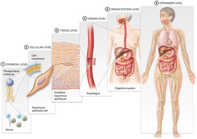
Hierarchy of Structural Organization
The human body is organized into a hierarchy of structural levels, from the smallest chemical components to the entire organism.
Chemical level: Atoms and molecules.
Cellular level: Cells and their organelles.
Tissue level: Groups of similar cells performing a function.
Organ level: Structures composed of two or more tissue types.
Organ system level: Groups of organs working together.
Organism level: The complete living being.
The 11 Organ Systems of the Human Body
The human body consists of eleven major organ systems, each with specific functions essential for life.
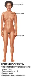
Integumentary system: Protects the body, produces vitamin D, retains water, regulates temperature.
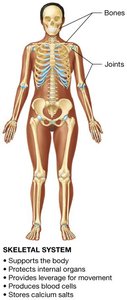
Skeletal system: Supports and protects, provides movement, stores minerals, produces blood cells.
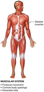
Muscular system: Produces movement, controls openings, generates heat.
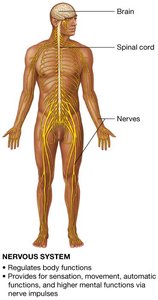
Nervous system: Regulates functions, provides sensation, movement, and higher mental functions.
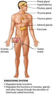
Endocrine system: Regulates functions via hormones.
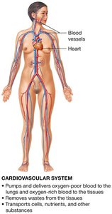
Cardiovascular system: Pumps blood, delivers oxygen, removes wastes, transports substances.
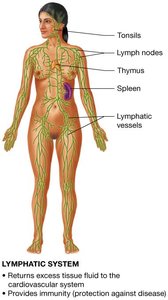
Lymphatic system: Returns fluid to blood, provides immunity.
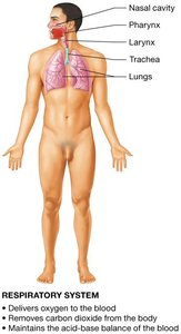
Respiratory system: Delivers oxygen, removes carbon dioxide, maintains acid-base balance.
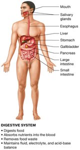
Digestive system: Digests food, absorbs nutrients, removes waste, maintains balance.
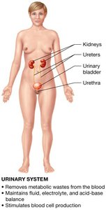
Urinary system: Removes wastes, maintains balance, stimulates blood cell production.
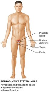
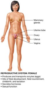
Reproductive system: Produces gametes, hormones, and enables reproduction.
Types of Anatomy and Physiology
Approaches to Studying the Human Body
Anatomy and physiology can be studied in several ways, each offering unique perspectives.
Systemic anatomy: Study by organ systems.
Regional anatomy: Study by body regions.
Surface anatomy: Study of surface markings.
Gross anatomy: Study of structures visible to the naked eye.
Microscopic anatomy: Study of structures visible only with a microscope (includes histology and cytology).
Physiology: Study of function, often by organ system or level of organization (e.g., neurophysiology, cardiovascular physiology).
The Language of Anatomy and Physiology
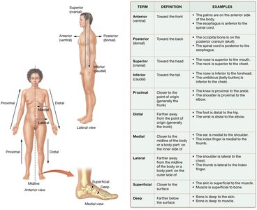
Anatomical Position
The anatomical position is a standardized frame of reference for describing the locations and relationships of body parts.
Body standing upright, feet shoulder-width apart, arms at sides, palms facing forward.
Terms "right" and "left" refer to the subject's sides, not the observer's.

Directional Terms
Directional terms are used to describe the locations of structures relative to other structures or locations in the body.
Anterior (ventral): Front of the body.
Posterior (dorsal): Back of the body.
Superior (cranial): Toward the head.
Inferior (caudal): Toward the tail.
Proximal: Closer to the point of origin.
Distal: Further from the point of origin.
Medial: Closer to the midline.
Lateral: Further from the midline.
Superficial: Closer to the surface.
Deep: Further from the surface.
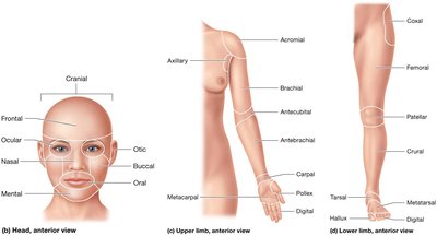
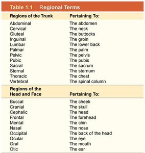
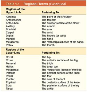
Regional Terms
Regional terms specify areas of the body, aiding in precise communication about location.
Regions of the trunk, head, face, upper limb, and lower limb are defined.
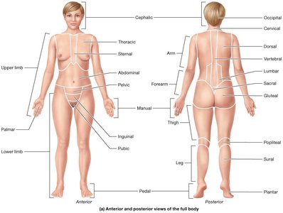
Region | Pertaining To |
|---|---|
Abdominal | The abdomen |
Cervical | The neck |
Gluteal | The buttocks |
Inguinal | The groin |
Lumbar | The lower back |
Palmar | The palm |
Pelvic | The pelvis |
Pubic | The pubis |
Sacral | The sacrum |
Sternal | The sternum |
Thoracic | The chest |
Vertebral | The spinal column |
Region | Pertaining To |
|---|---|
Buccal | The cheek |
Cranial | The head |
Cephalic | The head |
Frontal | The forehead |
Mental | The chin |
Nasal | The nose |
Occipital | The back of the head |
Ocular | The eye |
Oral | The mouth |
Otic | The ear |
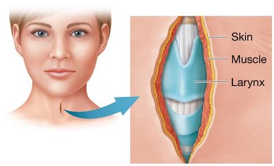
Concept Boost: Using Anatomical Terms
Combining regional and directional terms allows precise description of locations and procedures.
Example: Incision on left anterior cervical region, 1 cm lateral to midline, extending from 1 cm inferior to mental region to 2 cm superior to thoracic region, deep to skin and muscle but superficial to larynx.
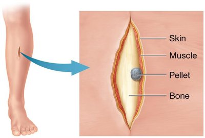
Example: Wound on left anteromedial crural region, 6 cm proximal to tarsal region and 10 cm distal to patellar region, pellet lodged deep to skin and muscle but superficial to bone.
Planes of Section
Planes of section are used to study the form and function of body regions by dividing the body or a body part.
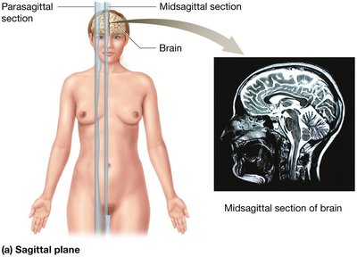
Sagittal plane: Divides body into right and left sections. Midsagittal: Equal halves; Parasagittal: Unequal halves.
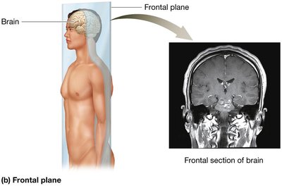
Frontal (coronal) plane: Divides body into anterior and posterior sections.
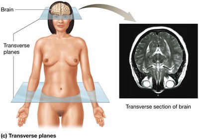
Transverse (horizontal) plane: Divides body into superior and inferior sections.
Oblique plane: Divides body at an angle.
Organization of the Human Body
Body Cavities
Body cavities are spaces within the body that house organs and provide protection.
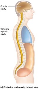
Posterior body cavity: Includes cranial cavity (brain) and vertebral cavity (spinal cord), filled with cerebrospinal fluid.
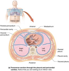
Anterior body cavity: Divided by diaphragm into thoracic and abdominopelvic cavities.
Thoracic cavity: Contains pleural cavities (lungs), mediastinum (heart, vessels, trachea, esophagus), and pericardial cavity (heart).

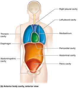
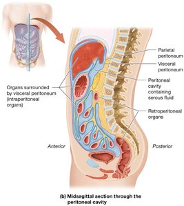
Abdominopelvic cavity: Contains abdominal and pelvic cavities, organs from digestive, lymphatic, reproductive, and urinary systems.
Peritoneal cavity: Subcavity within serous membrane.

Abdominopelvic Quadrants and Regions
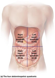
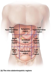
The abdominopelvic cavity can be divided into quadrants or nine regions for clinical and anatomical reference.
Quadrants: Right upper (RUQ), right lower (RLQ), left upper (LUQ), left lower (LLQ).
Regions: Right/left hypochondriac, epigastric, right/left lumbar, umbilical, right/left iliac, hypogastric.
Serous Membranes and Body Cavities
Serous membranes are thin sheets of tissue that form cavities and surround organs, providing lubrication and reducing friction.
Visceral layer: In contact with the organ.
Parietal layer: Attached to surrounding structures.
Serous fluid: Lubricates and prevents friction.
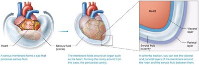
Serous Body Cavities
Three main serous membranes form body cavities:
Pleural membranes: Surround lungs; pleural cavity is the space between.
Pericardial membranes: Surround heart; pericardial cavity is the space between.
Peritoneal membranes: Surround some abdominal organs; peritoneal cavity is the space between. Retroperitoneal organs lie outside the peritoneal cavity.
Core Principles in Anatomy and Physiology
Homeostasis
Homeostasis is the maintenance of a stable internal environment. Physiological processes operate to keep variables within a narrow range.
Imbalance: Can lead to disease or death.
Variables: Include temperature, chemical composition, etc.
Core Principles
Feedback loops: Mechanisms that maintain homeostasis.
Structure and function: Form follows function at all levels.
Gradients: Differences in temperature, concentration, or pressure drive physiological processes.
Cell-to-cell communication: Coordination via chemical messengers or electrical signals.
Feedback Loops
Feedback loops are vital for homeostasis. Negative feedback opposes changes, while positive feedback amplifies them.
Negative feedback: Returns variable to normal range; ends when balance is restored.
Positive feedback: Amplifies change until a specific outcome is achieved (e.g., childbirth).
Structure and Function
The principle of complementarity states that the form of a structure is always suited to its function.
Gradients
Gradients are differences in temperature, concentration, or pressure between two connected regions. They drive many physiological processes.
Temperature gradient: Difference in temperature.
Concentration gradient: Difference in concentration.
Pressure gradient: Difference in pressure.
Cell-Cell Communication
Cells communicate to coordinate body functions and maintain homeostasis, using chemical messengers or electrical signals.
Additional info: These notes expand on brief points from the original slides, providing definitions, examples, and context for each concept. Tables are recreated for regional terms, and only images directly relevant to each topic are included.
