 Back
BackChapter 1: Introduction to Anatomy and Physiology – Structured Study Notes
Study Guide - Smart Notes
 Tailored notes based on your materials, expanded with key definitions, examples, and context.
Introduction to Anatomy and Physiology
Why Study Anatomy and Physiology?
Anatomy and physiology are foundational sciences for all health professions and biology-based careers. Understanding these subjects helps individuals make informed health decisions, interpret medical news, and become more knowledgeable about their own bodies.
Anatomy: The study of internal and external body structures and their physical relationships among other body parts.
Physiology: The study of how living organisms perform their vital functions.
Structure and function are closely related; anatomical features often determine physiological roles.
Levels of Organization in the Human Body
Chemical Level
The chemical level is the simplest level of organization, involving atoms and molecules that form the building blocks of matter.
Atoms: The smallest stable units of matter.
Molecules: Combinations of two or more atoms (e.g., H2O).
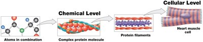
Cellular Level
Cells are the smallest living units in the human body. Humans are composed of eukaryotic cells, which contain organelles.
Cells: Basic units of structure and function in living organisms.
Tissue Level
Tissues are groups of similar cells that work together to perform specific functions. Most tissues contain more than one cell type.
Tissue: A group of cells with a common function.
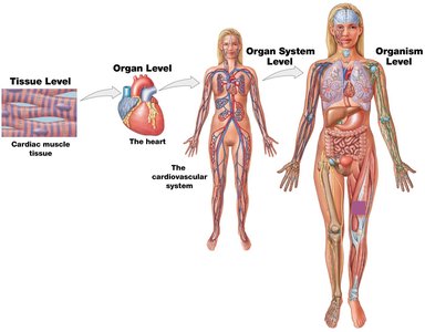
Organ Level
An organ is a distinct body structure composed of two or more tissue types that performs specific functions.
Organ: Structure with specialized functions (e.g., heart, liver).
Organ System Level
An organ system is a group of organs that interact to perform specific functions essential for the body’s survival.
Organ system: Multiple organs working together (e.g., cardiovascular system).
Organism Level
The organism level is the highest level of organization, representing the complete living individual.
Organism: An individual living entity (e.g., a human).
Organ Systems of the Human Body
Overview of Organ Systems
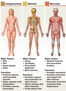
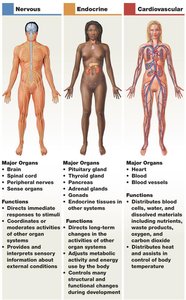
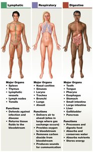
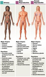
The human body is organized into eleven major organ systems, each with specific functions and major organs.
Integumentary: Skin, hair, nails – protection, temperature regulation.
Skeletal: Bones, cartilage – support, protection, blood cell formation.
Muscular: Skeletal muscles – movement, heat production.
Nervous: Brain, spinal cord – immediate response to stimuli.
Endocrine: Glands – long-term changes, metabolic activity.
Cardiovascular: Heart, blood vessels – transport of blood and nutrients.
Lymphatic: Spleen, lymph nodes – defense against infection.
Respiratory: Lungs, trachea – gas exchange.
Digestive: Stomach, intestines – nutrient absorption.
Urinary: Kidneys, bladder – waste elimination, water balance.
Reproductive: Ovaries/testes – production of sex cells, offspring.
Anatomical Terminology
Anatomical Position
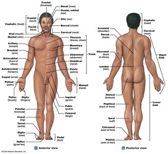
The anatomical position is the standard reference for describing body locations and directions. The body stands upright, feet together, arms at the sides, and palms facing forward.
Reduces confusion in anatomical descriptions.
Anterior (front) and posterior (back) views are used for reference.
Directional Terms
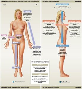
Directional terms describe the locations of structures relative to other structures or locations in the body.
Anterior (ventral): Toward the front
Posterior (dorsal): Toward the back
Superior (cranial): Toward the head
Inferior (caudal): Toward the feet
Lateral: Away from the midline
Medial: Toward the midline
Proximal: Closer to the point of attachment
Distal: Farther from the point of attachment
Superficial: Near the surface
Deep: Farther from the surface
Prone: Face-down
Supine: Face-up

Sectional Anatomy
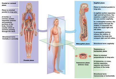
Body Planes and Sections
Sectional anatomy involves dividing the body into planes to study its internal structures. Medical imaging often uses these planes for diagnostic purposes.
Frontal (coronal) plane: Divides the body into anterior and posterior portions.
Sagittal plane: Divides the body into right and left portions. The midsagittal plane is exactly in the middle; the parasagittal plane is offset.
Transverse (horizontal) plane: Divides the body into superior and inferior portions.
Body Cavities
Major Body Cavities
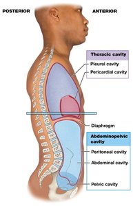
Body cavities are closed, fluid-filled spaces lined by serous membranes. They protect organs and allow for changes in organ size and shape.
Thoracic cavity: Contains pleural cavities (lungs) and pericardial cavity (heart), separated from the abdominopelvic cavity by the diaphragm.
Abdominopelvic cavity: Contains abdominal and pelvic organs, separated from the thoracic cavity by the diaphragm.
Serous Membranes
Serous membranes line body cavities and cover organs. They consist of two layers:
Visceral layer: Covers the organ.
Parietal layer: Lines the cavity wall.
Serous fluid: Reduces friction between layers during organ movement.

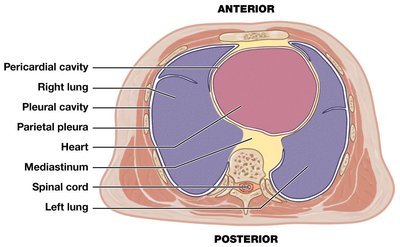
Thoracic Cavity Subdivisions
The thoracic cavity includes the pleural cavities (lungs), pericardial cavity (heart), and the mediastinum (connective tissue mass containing the heart, trachea, esophagus, and major vessels).
Pleura: Serous membrane of the pleural cavity.
Serous pericardium: Serous membrane of the pericardial cavity.
Abdominopelvic Cavity Subdivisions
The abdominopelvic cavity is divided into the abdominal cavity (liver, stomach, spleen, intestines) and pelvic cavity (urinary bladder, reproductive organs, distal large intestine). The peritoneum is the serous membrane lining this cavity.
Peritoneum: Parietal peritoneum lines the body wall; visceral peritoneum covers organs.
Retroperitoneal organs: Located behind the peritoneum (e.g., kidneys, pancreas).
Abdominopelvic Quadrants and Regions
Clinicians use quadrants and regions to describe locations of pain or injury in the abdomen.
Quadrants: Right upper, left upper, right lower, left lower.
Regions: More precise anatomical divisions used in research and clinical settings.
Homeostasis
Definition and Importance
Homeostasis is the maintenance of a stable internal environment through continuous physiological processes. It is essential for survival and involves the regulation of variables such as temperature and blood pressure.
Homeostatic regulation: Adjustment of physiological systems to maintain stability.
Three main components: Receptor (detects change), Control center (processes information), Effector (carries out response).
Set Point
Each physiological variable has a set point or normal range that the body attempts to maintain (e.g., body temperature, blood pressure).
Deviation from the set point triggers regulatory mechanisms.
Negative Feedback
Negative feedback mechanisms oppose deviations from the set point, restoring physiological variables to normal ranges. This is the most common type of homeostatic regulation.
Example: Thermoregulation – if body temperature rises or falls, effectors act to reverse the change.
Positive Feedback
Positive feedback mechanisms enhance or intensify the original stimulus. These are less common and typically occur when a rapid, definitive outcome is needed.
Examples: Blood clotting, labor and delivery.
