 Back
BackChapter 1: Introduction to Anatomy & Physiology – Study Notes
Study Guide - Smart Notes
 Tailored notes based on your materials, expanded with key definitions, examples, and context.
Introduction to Anatomy & Physiology
Using the Text and Art Effectively
Understanding anatomy and physiology requires integrating textual information with visual aids. The textbook is structured to build knowledge progressively, and figures are placed near relevant text for clarity.
Strategy 1: Read the text, then study the corresponding image to reinforce understanding.
Strategy 2: Focus on learning outcomes, as they highlight essential knowledge and skills for assessment.
Strategy 3: Engage with your instructor, follow the syllabus, keep up with readings, and utilize available study tools.

Defining Anatomy and Physiology
Basic Definitions and Relationship
Anatomy is the study of body structures, both internal and external, and their relationships. Physiology is the study of how living organisms perform vital functions. These disciplines are closely linked by the principle of complementarity: structure determines function, and function reflects structure.
Gross (Macroscopic) Anatomy: Study of large, visible structures.
Microscopic Anatomy: Study of structures requiring magnification (cells, tissues).
Specialties in Anatomy: Surface, regional, sectional, systemic, clinical, developmental, and embryology.
Specialties in Physiology: Cell, organ, systemic, and pathological physiology.
Physicians use anatomical and physiological knowledge, along with chemical and psychological data, to diagnose patients using the scientific method.
Levels of Organization in the Human Body
Hierarchy from Atoms to Organism
The human body is organized into six hierarchical levels, each building on the previous:
Chemical Level: Atoms and molecules (e.g., proteins, DNA).
Cellular Level: Cells, the smallest living units.
Tissue Level: Groups of similar cells performing specific functions.
Organ Level: Two or more tissues working together (e.g., heart).
Organ System Level: Groups of organs performing related functions (e.g., cardiovascular system).
Organism Level: The complete living individual.
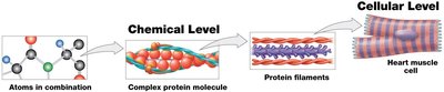
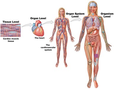
Major Organ Systems
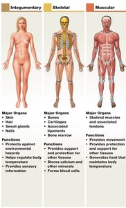
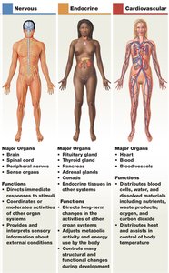
The human body contains 11 organ systems, each with specific organs and functions:
Integumentary (skin, hair, nails): Protection, temperature regulation.
Skeletal (bones, cartilage): Support, protection, blood formation.
Muscular (muscles, tendons): Movement, heat production.
Nervous (brain, spinal cord): Immediate response, coordination.
Endocrine (glands): Long-term changes, hormone production.
Cardiovascular (heart, blood vessels): Transport of blood, nutrients, and waste.
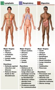
Lymphatic (spleen, lymph nodes): Defense, fluid return.
Respiratory (lungs, trachea): Gas exchange.
Digestive (stomach, intestines): Nutrient absorption, waste elimination.
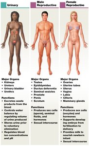
Urinary (kidneys, bladder): Waste excretion, water balance.
Reproductive (testes, ovaries): Production of sex cells, offspring.
Medical Terminology
Origins and Importance
Medical terminology is based on word roots, prefixes, suffixes, and combining forms. Understanding these components aids in learning anatomy and physiology. Standardized terms reduce confusion, though some eponyms (names based on discoverers) are still used alongside precise terms.
Anatomical Terminology
Body Regions, Sections, and Positions
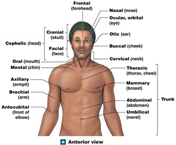
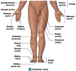
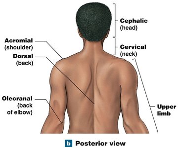
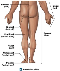
Surface anatomy identifies structures on or near the body surface using anatomical landmarks. The anatomical position is the standard reference: standing, hands at sides, palms forward, feet together.
Anterior (front) and Posterior (back) views are used for orientation.
Supine: Lying face up; Prone: Lying face down.
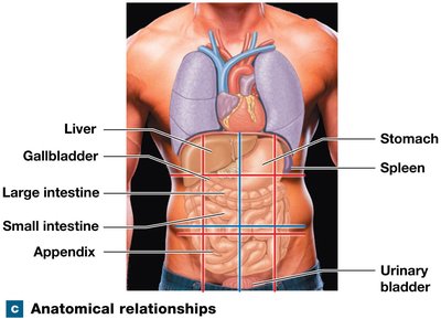
Abdominopelvic Quadrants and Regions
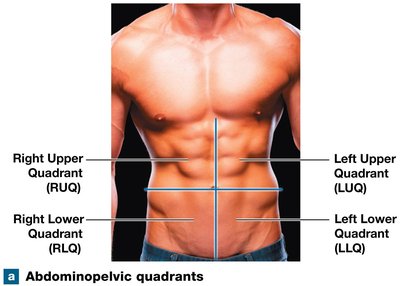
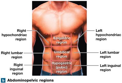
The abdominopelvic area is divided for clinical and anatomical reference:
Quadrants: Right Upper (RUQ), Left Upper (LUQ), Right Lower (RLQ), Left Lower (LLQ).
Regions: Nine regions for more precise localization (e.g., epigastric, umbilical, hypogastric).
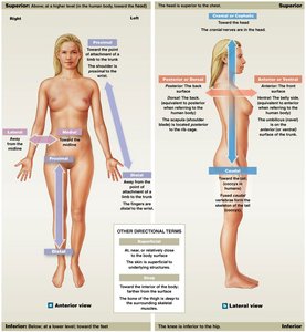
Directional References
Directional terms describe the location of structures relative to each other (e.g., superior, inferior, medial, lateral, proximal, distal).
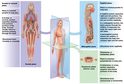
Sectional Anatomy and Planes
Sections and planes are used to visualize internal structures:
Frontal (coronal) plane: Divides body into anterior and posterior parts.
Sagittal plane: Divides body into left and right parts (midsagittal = equal halves; parasagittal = unequal).
Transverse (horizontal) plane: Divides body into superior and inferior parts (cross section).
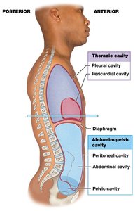
Body Cavities
Major Cavities and Their Functions
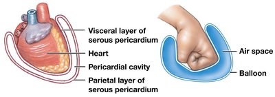
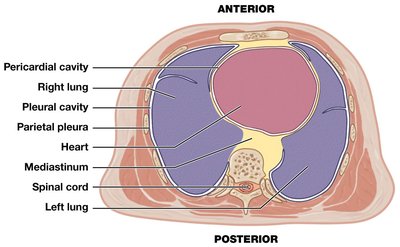
Body cavities are closed, fluid-filled spaces lined by serous membranes, housing and protecting vital organs (viscera). They allow organs to change size and shape and protect them from shocks.
Serous membrane (serosa): Has parietal (lines cavity) and visceral (covers organ) layers, with serous fluid reducing friction.
Thoracic and Abdominopelvic Cavities
Thoracic cavity: Contains right and left pleural cavities (lungs), pericardial cavity (heart), and mediastinum (central tissue mass).
Abdominopelvic cavity: Contains abdominal (digestive organs) and pelvic (reproductive, rectum, bladder) cavities, and the peritoneal cavity (lined by peritoneum).
Retroperitoneal space: Area behind the peritoneum (e.g., kidneys, pancreas).
Infraperitoneal: Organs below the peritoneal cavity (e.g., bladder).
Homeostasis
Definition and Mechanisms
Homeostasis is the maintenance of a stable internal environment through continuous physiological processes. Systems respond to internal and external changes to keep variables (e.g., temperature, blood pressure) within normal ranges.
Autoregulation: Local, automatic response to environmental change.
Extrinsic regulation: Controlled by nervous (fast, short-term) or endocrine (slow, long-term) systems.
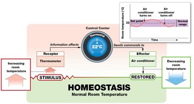
A homeostatic regulatory mechanism includes:
Receptor: Detects stimulus.
Control center: Processes information and sends commands.
Effector: Carries out commands to restore balance.
Negative and Positive Feedback
Types of Homeostatic Regulation
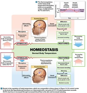
Negative feedback: Opposes deviations from normal; effector response negates the original stimulus, maintaining stability (e.g., body temperature regulation).
Positive feedback: Enhances deviations; response amplifies the original change, used for rapid completion of processes (e.g., blood clotting).
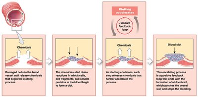
Systems Integration and Dynamic Equilibrium
Organ systems work together to maintain homeostasis. Adjustments in one system affect others. Homeostasis is a dynamic equilibrium, with physiological systems continually adapting. Failure to maintain homeostasis leads to disease or death.
Table: Roles of Organ Systems in Homeostatic Regulation
The following table summarizes how different organ systems contribute to homeostatic regulation:
Internal Stimulus | Primary Organ Systems Involved | Functions of the Organ Systems |
|---|---|---|
Body temperature | Integumentary, Muscular, Cardiovascular, Nervous | Heat loss, heat production, heat distribution, coordination of blood flow and temperature regulation |
Nutrient concentration | Digestive, Cardiovascular, Urinary, Skeletal | Nutrient absorption, storage, release, distribution, and control of nutrient loss |
Oxygen, carbon dioxide levels | Respiratory, Cardiovascular | Absorption of oxygen, elimination of carbon dioxide, internal transport |
Levels of toxins and pathogens | Lymphatic | Removal, destruction, or inactivation of toxins and pathogens |
Body fluid volume | Urinary, Digestive, Integumentary, Cardiovascular, Lymphatic | Elimination/conservation of water, absorption, loss through perspiration, distribution |
Waste concentration | Urinary, Digestive, Cardiovascular | Excretion of wastes, elimination in feces, transport to excretion sites |
Blood pressure | Cardiovascular, Nervous, Endocrine | Pressure generation, adjustments in heart rate and vessel diameter |
