 Back
BackChapter 1: The Human Body – An Orientation (Anatomy & Physiology Study Notes)
Study Guide - Smart Notes
 Tailored notes based on your materials, expanded with key definitions, examples, and context.
Form Determines Function
Introduction to Anatomy and Physiology
The study of the human body is divided into anatomy (structure) and physiology (function). These disciplines are inseparable, as the function of a body part always reflects its structure—a concept known as the principle of complementarity of structure and function.
Anatomy: Study of the structure of body parts and their relationships to one another.
Physiology: Study of the function of body parts and how they carry out life-sustaining activities.
Key Concept: What a structure can do depends on its specific form.
Topics and Subdivisions of Anatomy
Macroscopic and Microscopic Anatomy
Gross (Macroscopic) Anatomy: Study of large, visible structures.
Regional Anatomy: Examines all structures in a particular area of the body.
System Anatomy: Focuses on one organ system (e.g., cardiovascular, nervous).
Surface Anatomy: Studies internal structures as they relate to the overlying skin.
Microscopic Anatomy: Structures too small to be seen with the naked eye.
Cytology: Study of cells.
Histology: Study of tissues.
Developmental Anatomy: Study of anatomical and physiological development throughout life.
Embryology: Study of developments before birth.
Topics and Subdivisions of Physiology
Organ System Physiology
Physiology is based on organ systems (e.g., renal, cardiovascular physiology).
Focuses on cellular and molecular levels, emphasizing chemical reactions in cells.
Requires understanding of basic physical and chemical principles.
Levels of Structural Organization
Hierarchy of the Human Body
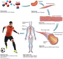
The human body is organized from the smallest chemical level to the entire organism.
Chemical Level: Atoms combine to form molecules.
Cellular Level: Cells are made up of molecules and organelles.
Tissue Level: Tissues consist of similar types of cells.
Organ Level: Organs are made up of different types of tissues.
Organ System Level: Organ systems consist of different organs that work together.
Organismal Level: The human organism is made up of many organ systems.
Requirements for Life
Necessary Life Functions
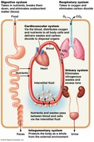
Humans are multicellular organisms whose bodily functions are distributed among 11 organ systems. These systems work together to maintain life.
Maintaining Boundaries: Separation between internal and external environments (e.g., plasma membranes, skin).
Movement: Muscular system allows movement; skeletal system provides framework.
Responsiveness: Ability to sense and respond to stimuli (e.g., withdrawal reflex, breathing rate).
Digestion: Breakdown of ingested food and absorption of nutrients.
Metabolism: All chemical reactions in body cells, including catabolism (breakdown) and anabolism (synthesis).
Excretion: Removal of wastes from metabolism and digestion.
Reproduction: Cellular division for growth/repair and production of offspring.
Growth: Increase in size of a body part or organism.
Survival Needs
Nutrients: Chemicals for energy and cell building (carbohydrates, proteins, fats, vitamins, minerals).
Oxygen: Essential for cellular respiration and energy release from food.
Water: Most abundant chemical in the body; necessary for chemical reactions.
Normal Body Temperature: Needed for proper rates of chemical reactions (about 37°C).
Appropriate Atmospheric Pressure: Required for adequate breathing and gas exchange.
Organ Systems Overview
The 11 Organ Systems
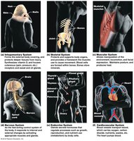
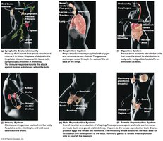
The human body contains 11 organ systems, each with specific functions essential for survival.
Integumentary System
Skeletal System
Muscular System
Nervous System
Endocrine System
Cardiovascular System
Lymphatic System
Respiratory System
Digestive System
Urinary System
Reproductive System
Homeostasis
Maintaining Internal Balance
Homeostasis is the maintenance of relatively stable internal conditions despite continuous changes in the environment. It is a dynamic state of equilibrium, maintained by all organ systems, especially the nervous and endocrine systems.
Variable: A factor or event being regulated (e.g., body temperature, blood sugar).
Homeostatic Control Mechanisms
Homeostatic control involves three main components:
Receptor (Sensor): Monitors the environment and responds to stimuli by sending information to the control center via the afferent pathway.
Control Center: Determines the set point, analyzes input, and determines the appropriate response.
Effector: Receives output from the control center via the efferent pathway and provides the means to respond, either reducing (negative feedback) or enhancing (positive feedback) the stimulus.
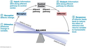
Negative Feedback
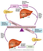
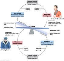
Negative feedback mechanisms reduce or shut off the original stimulus, causing the variable to change in the opposite direction of the initial change. Most homeostatic controls are negative feedback mechanisms.
Examples: Regulation of body temperature, regulation of blood glucose by insulin.
Positive Feedback
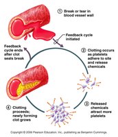
Positive feedback mechanisms enhance or exaggerate the original stimulus, causing the variable to continue in the same direction. These mechanisms usually control infrequent events that do not require continuous adjustment.
Examples: Enhancement of labor contractions by oxytocin, platelet plug formation and blood clotting.
Homeostatic Imbalance
Most diseases result from a disturbance in homeostasis, known as homeostatic imbalance. Aging and overwhelmed negative feedback mechanisms can lead to destructive positive feedback, such as heart failure.
Anatomical Terms
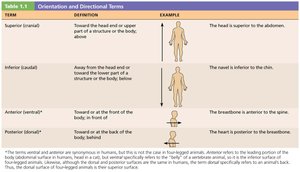
Standard Anatomical Position and Directional Terms
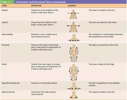
The standard anatomical position is body erect, feet slightly apart, palms facing forward, and thumbs pointing away from the body. Directional terms describe the location of one body structure relative to another, always based on the standard anatomical position.
Term | Definition | Example |
|---|---|---|
Superior (cranial) | Toward the head end or upper part of a structure or the body; above | The head is superior to the abdomen. |
Inferior (caudal) | Away from the head end or toward the lower part of a structure or the body; below | The navel is inferior to the chin. |
Anterior (ventral) | Toward or at the front of the body; in front of | The breastbone is anterior to the spine. |
Posterior (dorsal) | Toward or at the back of the body; behind | The heart is posterior to the breastbone. |
Term | Definition | Example |
|---|---|---|
Medial | Toward or at the midline of the body; on the inner side of | The heart is medial to the arm. |
Lateral | Away from the midline of the body; on the outer side of | The arms are lateral to the chest. |
Intermediate | Between a more medial and a more lateral structure | The collarbone is intermediate between the breastbone and shoulder. |
Proximal | Closer to the origin of the body part or the point of attachment of a limb to the body trunk | The elbow is proximal to the wrist. |
Distal | Farther from the origin of a body part or the point of attachment of a limb to the body trunk | The knee is distal to the thigh. |
Superficial (external) | Toward or at the body surface | The skin is superficial to the skeletal muscles. |
Deep (internal) | Away from the body surface; more internal | The lungs are deep to the skin. |
Regional Terms
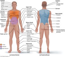
The body is divided into two major regions: axial (head, neck, trunk) and appendicular (limbs). Regional terms designate specific areas within these divisions.
Body Planes and Sections
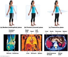
Major Body Planes
Sagittal Plane: Divides the body vertically into right and left parts.
Midsagittal (Median) Plane: Cut made perfectly on the midline.
Parasagittal Plane: Cut off-centered, not on the midline.
Frontal (Coronal) Plane: Divides the body vertically into anterior and posterior parts.
Transverse (Horizontal) Plane: Divides the body horizontally into superior and inferior parts; produces a cross-section.
Oblique Section: Cuts made at an angle other than 90° to the vertical plane.
Body Cavities and Membranes
Major Body Cavities
The body contains internal cavities that are closed to the environment, providing protection to organs.
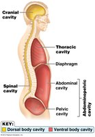
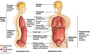
Dorsal Body Cavity: Protects the nervous system; includes cranial and vertebral (spinal) cavities.
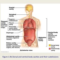
Ventral Body Cavity: Houses internal organs (viscera); includes thoracic and abdominopelvic cavities, separated by the diaphragm.
Ventral Body Cavity Subdivisions
Thoracic Cavity: Surrounded by ribs and chest muscles; contains pleural cavities (lungs), mediastinum (heart, trachea, esophagus), and pericardial cavity (heart).
Abdominopelvic Cavity: Inferior to the thoracic cavity; contains abdominal cavity (digestive organs) and pelvic cavity (urinary bladder, reproductive organs, rectum).
Serous Membranes
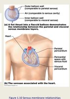
Serous membranes (serosa) are thin, double-layered membranes covering surfaces in the ventral body cavity. They are named for the specific cavity and organs they are associated with:
Parietal Serosa: Lines internal body cavity walls.
Visceral Serosa: Covers internal organs.
Serous Fluid: Fills the slit-like cavity between layers, reducing friction.
Pericardium: Heart
Pleurae: Lungs
Peritoneum: Abdominopelvic cavity
Abdominopelvic Divisions
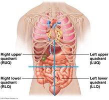
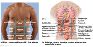
The abdominopelvic cavity is divided into quadrants and regions for clinical and anatomical reference.
Quadrants: Right upper (RUQ), left upper (LUQ), right lower (RLQ), left lower (LLQ).
Regions: Right hypochondriac, epigastric, left hypochondriac, right lumbar, umbilical, left lumbar, right iliac (inguinal), hypogastric, left iliac (inguinal).
Other Body Cavities
Exposed to Environment: Oral and digestive, nasal, orbital, middle ear cavities.
Not Exposed: Synovial (joint) cavities.
Homeostatic Imbalances of Serous Membranes
Inflammation of serous membranes can cause pain and dysfunction (e.g., pleurisy, peritonitis).
