 Back
BackChapter 1: The Human Body – An Orientation
Study Guide - Smart Notes
 Tailored notes based on your materials, expanded with key definitions, examples, and context.
Chapter 1: The Human Body – An Orientation
Introduction to Human Anatomy and Physiology
This chapter introduces foundational concepts and terminology essential for the study of human anatomy and physiology. Understanding these basics is crucial for success in health-related fields and for effective communication in clinical settings.
Anatomy is the study of the structure of body parts and their relationships to one another.
Physiology is the study of the function of the body’s structural machinery.
Both disciplines are closely linked, as structure often determines function.
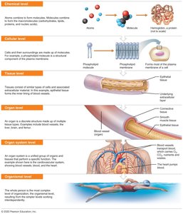
Levels of Structural Organization
The human body is organized into a hierarchy of structural levels, each building upon the previous one. Understanding these levels helps explain how complex functions arise from simpler components.
Chemical Level: Atoms combine to form molecules, which are the building blocks of cells.
Cellular Level: Cells are the smallest living units, composed of organelles and surrounded by a plasma membrane.
Tissue Level: Tissues are groups of similar cells that perform a common function (e.g., muscle tissue, nervous tissue).
Organ Level: Organs are structures composed of at least two types of tissues working together for a specific function (e.g., heart, liver).
Organ System Level: Organ systems consist of different organs that work closely together (e.g., digestive system, respiratory system).
Organismal Level: The human organism is made up of many organ systems functioning together.
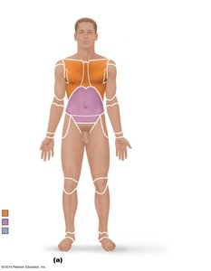
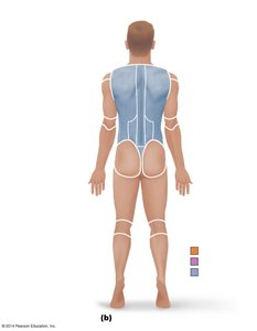
Anatomical Position and Regional Terms
Standard anatomical position and regional terminology provide a universal reference for describing locations and relationships of body parts.
Anatomical Position: The body stands erect, facing forward, arms at the sides with palms facing forward, and feet slightly apart.
Axial Region: Includes the head, neck, and trunk.
Appendicular Region: Includes the limbs (arms and legs).
Specific regional terms (e.g., cephalic for head, brachial for arm) are used to describe precise locations.
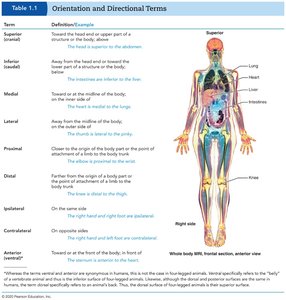
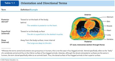
Orientation and Directional Terms
Directional terms are used to explain where one body structure is in relation to another. These terms are essential for clear communication in anatomy and clinical practice.
Superior (cranial): Toward the head or upper part of a structure.
Inferior (caudal): Away from the head or toward the lower part.
Anterior (ventral): Toward the front of the body.
Posterior (dorsal): Toward the back of the body.
Medial: Toward the midline of the body.
Lateral: Away from the midline.
Proximal: Closer to the origin of a body part or point of attachment.
Distal: Farther from the origin or point of attachment.
Superficial (external): Toward or at the body surface.
Deep (internal): Away from the body surface; more internal.
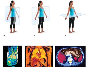
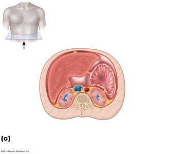
Body Planes and Sections
Body planes are imaginary lines used to divide the body into sections for anatomical study and medical imaging.
Median (midsagittal) plane: Divides the body into equal right and left halves.
Frontal (coronal) plane: Divides the body into anterior and posterior parts.
Transverse (horizontal) plane: Divides the body into superior and inferior parts.
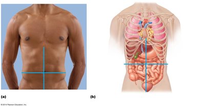
Abdominopelvic Quadrants
The abdominopelvic cavity is divided into four quadrants to help localize organs and describe pain or injury locations.
Right Upper Quadrant (RUQ)
Left Upper Quadrant (LUQ)
Right Lower Quadrant (RLQ)
Left Lower Quadrant (LLQ)
Each quadrant contains specific organs, aiding in clinical diagnosis.
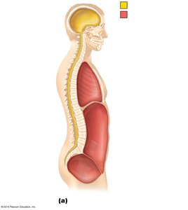
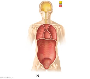
Body Cavities and Membranes
The body contains several major cavities that house and protect internal organs. These cavities are lined by membranes that provide lubrication and reduce friction.
Dorsal Body Cavity: Includes the cranial cavity (brain) and vertebral cavity (spinal cord).
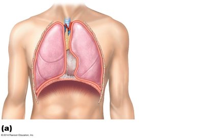
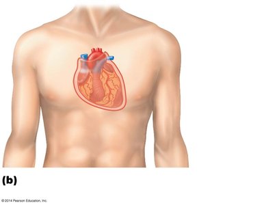
Ventral Body Cavity: Includes the thoracic cavity (heart and lungs) and abdominopelvic cavity (digestive, urinary, and reproductive organs).
Serous Membranes: Double-layered membranes that line body cavities and cover organs. Types include pleura (lungs), pericardium (heart), and peritoneum (abdominal organs).
Medical Imaging Techniques
Modern imaging techniques allow visualization of internal structures for diagnosis and treatment. Each method has unique advantages and applications.
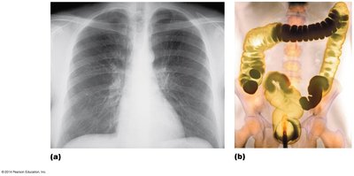
X-ray: Uses radiation to view dense structures like bones and some organs.
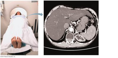
Computed Tomography (CT): Produces cross-sectional images using X-rays and computer processing.
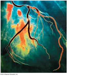
Digital Subtraction Angiography (DSA): Visualizes blood vessels by subtracting pre-contrast images from post-contrast images.
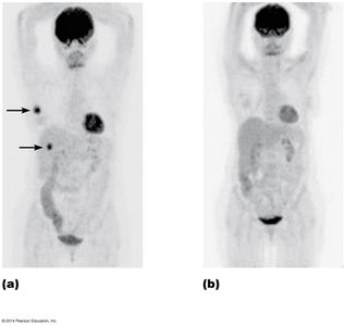
Positron Emission Tomography (PET): Detects metabolic activity using radioactive tracers.
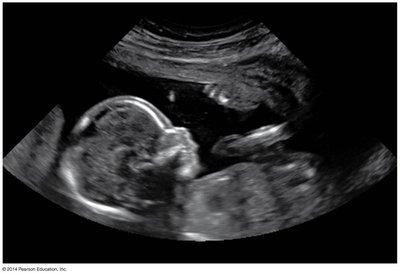
Ultrasound: Uses high-frequency sound waves to image soft tissues, commonly used in obstetrics.
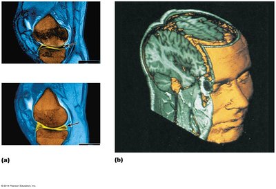
Magnetic Resonance Imaging (MRI): Uses magnetic fields and radio waves to produce detailed images of soft tissues.
Summary Table: Orientation and Directional Terms
The following table summarizes key orientation and directional terms used in human anatomy:
Term | Definition/Example |
|---|---|
Superior (cranial) | Toward the head or upper part of a structure (e.g., the head is superior to the abdomen). |
Inferior (caudal) | Away from the head or toward the lower part (e.g., the navel is inferior to the chin). |
Anterior (ventral) | Toward the front of the body (e.g., the breastbone is anterior to the spine). |
Posterior (dorsal) | Toward the back of the body (e.g., the heart is posterior to the sternum). |
Medial | Toward the midline of the body (e.g., the heart is medial to the arm). |
Lateral | Away from the midline (e.g., the arms are lateral to the chest). |
Proximal | Closer to the origin of a body part (e.g., the elbow is proximal to the wrist). |
Distal | Farther from the origin (e.g., the knee is distal to the thigh). |
Superficial (external) | Toward or at the body surface (e.g., the skin is superficial to the muscles). |
Deep (internal) | Away from the body surface; more internal (e.g., the lungs are deep to the skin). |
Clinical Application Example
Scenario: A trauma patient presents with pain in the left lower quadrant after a car accident. Organs at risk include the descending colon, sigmoid colon, and left ovary (in females). Imaging such as CT or ultrasound may be ordered to assess injury.
Key Takeaways
Mastery of anatomical terminology and orientation is foundational for all health professions.
Understanding body planes, regions, and cavities is essential for interpreting medical images and clinical scenarios.
Modern imaging techniques provide powerful tools for non-invasive diagnosis and treatment planning.
