 Back
BackChapter 1: The Human Body – An Orientation
Study Guide - Smart Notes
 Tailored notes based on your materials, expanded with key definitions, examples, and context.
Overview of Anatomy and Physiology
Definitions and Scope
Anatomy is the study of the structure of body parts and their relationships to one another. It is divided into:
Gross (macroscopic) anatomy: Study of structures visible to the naked eye.
Microscopic anatomy: Study of structures requiring a microscope.
Developmental anatomy: Study of structural changes throughout life, including embryology (development before birth).
Physiology is the study of the function of the body’s structural machinery, often at the cellular or molecular level. It includes:
Renal physiology: Kidney function
Neurophysiology: Nervous system function
Cardiovascular physiology: Heart and blood vessel function
Specialized branches include pathological anatomy (disease-related changes), radiographic anatomy (imaging), and molecular biology (subcellular structures).
Principle of Complementarity
Structure and Function Relationship
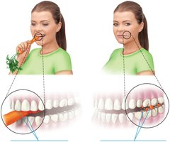
The principle of complementarity states that function always reflects structure; what a structure can do depends on its specific form.
Example: The sharp edges of incisors are ideal for cutting, while the flat surfaces of molars are suited for grinding.
Levels of Structural Organization
Hierarchy of Complexity
The human body is organized into a hierarchy of structural levels:
Chemical level: Atoms combine to form molecules.
Cellular level: Cells are made up of molecules.
Tissue level: Tissues consist of similar types of cells.
Organ level: Organs are made up of different types of tissues.
Organ system level: Organ systems consist of different organs that work together closely.
Organismal level: The human organism is made up of many organ systems.
Organ Systems of the Body
Major Organ Systems and Their Functions
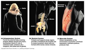
Integumentary System: Forms the external body covering, protects deeper tissues, synthesizes vitamin D, and houses cutaneous receptors.
Skeletal System: Protects and supports body organs, provides a framework for muscles, stores minerals.
Muscular System: Allows manipulation of the environment, locomotion, and facial expression; maintains posture; produces heat.
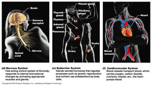
Nervous System: Fast-acting control system, responds to internal and external changes.
Endocrine System: Glands secrete hormones that regulate processes such as growth, reproduction, and metabolism.
Cardiovascular System: Blood vessels transport blood, which carries oxygen, nutrients, wastes, etc.; the heart pumps blood.
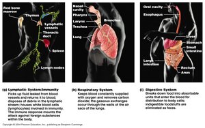
Lymphatic/Immune System: Picks up fluid leaked from blood vessels, houses white blood cells, mounts immune response.
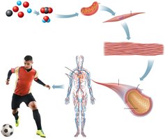
Respiratory System: Keeps blood supplied with oxygen and removes carbon dioxide.
Digestive System: Breaks down food into absorbable units, eliminates indigestible foodstuffs as feces.
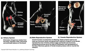
Urinary System: Eliminates nitrogenous wastes, regulates water, electrolyte, and acid-base balance.
Male and Female Reproductive Systems: Production of offspring; testes produce sperm and male sex hormones, ovaries produce eggs and female sex hormones.
Necessary Life Functions
Basic Functions Required for Life
Maintaining boundaries: Separation between internal and external environments (e.g., plasma membranes, skin).
Movement: Locomotion, propulsion, and contractility.
Responsiveness: Ability to sense and respond to stimuli.
Digestion: Breakdown of ingested foodstuffs.
Metabolism: All chemical reactions in the body, including catabolism (breakdown) and anabolism (synthesis).
Excretion: Removal of wastes.
Reproduction: Cellular (cell division) and organismal (offspring production).
Growth: Increase in size of a body part or organism.
Survival Needs
Essential Factors for Survival
Nutrients: Chemicals for energy and cell building.
Oxygen: Required for metabolic reactions.
Water: Environment for chemical reactions.
Normal body temperature: Necessary for proper metabolic rates.
Atmospheric pressure: Required for breathing and gas exchange.
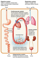
Organ Systems Interrelationships
Cooperation Among Systems
Organ systems work cooperatively to maintain life. All cells depend on organ systems to meet their survival needs.
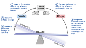
Homeostasis
Maintaining Internal Balance
Homeostasis is the maintenance of relatively stable internal conditions despite continuous environmental changes. It is a dynamic equilibrium maintained by all organ systems, primarily the nervous and endocrine systems.
Variables regulated include blood sugar, body temperature, calcium levels, heart rate, and breathing.
Homeostatic control involves three components: receptor (sensor), control center, and effector.
Homeostatic Control Mechanism
Receptor: Monitors environment and responds to stimuli.
Control Center: Determines set point and appropriate response.
Effector: Provides means to respond; response reduces (negative feedback) or enhances (positive feedback) the stimulus.
Feedback Mechanisms
Negative Feedback
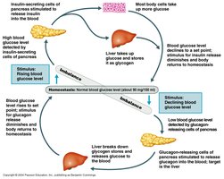
Most homeostatic control mechanisms are negative feedback, where the response reduces or shuts off the original stimulus, returning the variable to its set point.
Examples: Regulation of body temperature and blood glucose by insulin.
Positive Feedback
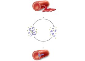
In positive feedback, the response enhances or exaggerates the original stimulus. This is less common but important in processes such as blood clotting and childbirth.
Example: Platelet plug formation during blood clotting.
Anatomical Position and Directional Terms
Standard Reference Position
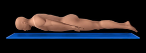
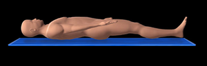
The anatomical position is the standard body position used as a reference in anatomy: body erect, feet slightly apart, palms facing forward, thumbs pointing away from the body. Other positions include prone (lying face down), supine (lying face up), and fetal position.

Orientation and Directional Terms
Directional terms describe the location of one body part relative to another. Examples include superior/inferior, anterior/posterior, medial/lateral, proximal/distal, superficial/deep.
Regional Terms
Body Regions
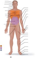
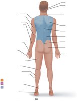
Regional terms specify areas within major body divisions (axial and appendicular). Examples include cephalic (head), cervical (neck), thoracic (chest), abdominal, pelvic, upper limb, lower limb, etc.
Body Planes and Sections
Dividing the Body for Study
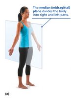
Sagittal plane: Divides body into right and left parts. Midsagittal is exactly in the midline; parasagittal is offset from the midline.
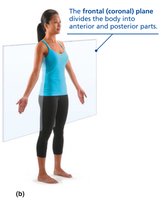
Frontal (coronal) plane: Divides body into anterior and posterior parts.
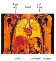
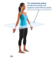
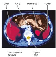
Transverse (horizontal) plane: Divides body into superior and inferior parts.
Oblique section: Cuts made at angles other than 90° to the vertical plane.
Body Cavities and Membranes
Major Body Cavities
Dorsal cavity: Protects the nervous system; includes cranial (brain) and vertebral (spinal cord) cavities.
Ventral cavity: Houses internal organs (viscera); includes thoracic and abdominopelvic cavities.
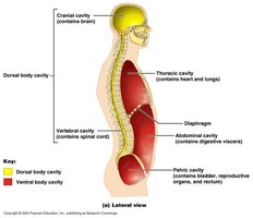
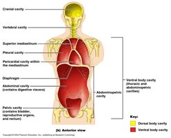
Thoracic and Abdominopelvic Cavities
Thoracic cavity: Subdivided into pleural cavities (lungs), mediastinum (heart, esophagus, trachea), and pericardial cavity (heart).
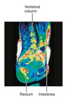
Abdominopelvic cavity: Subdivided into abdominal (digestive organs) and pelvic (bladder, reproductive organs, rectum) cavities.
Membranes in the Ventral Body Cavity
Serosa (serous membrane): Thin, double-layered membranes that cover surfaces in the ventral body cavity. The parietal serosa lines cavity walls; the visceral serosa covers organs. The cavity between is filled with lubricating serous fluid.
Pleura: Surrounds lungs
Pericardium: Surrounds heart
Peritoneum: Surrounds abdominopelvic organs
Dorsal Body Cavity Membranes
Meninges: Three layers of protective tissue (dura mater, arachnoid mater, pia mater) that line the cranial cavity and vertebral canal.
Abdominopelvic Regions and Quadrants
Nine Regions (Anatomists) and Four Quadrants (Clinicians)
Nine regions: right/left hypochondriac, epigastric, right/left lumbar, umbilical, right/left iliac (inguinal), hypogastric (pubic).
Four quadrants: right upper, left upper, right lower, left lower.
Clinical Assessment and Medical Imaging
Clinical Assessment Techniques
Inspection: Observing the body for changes
Palpation: Touching body surfaces
Auscultation: Listening to body sounds
Percussion: Tapping and listening to echoes
Medical Imaging Techniques
Radiography (X-rays): Visualizes internal structures; dense structures appear white.
Magnetic Resonance Imaging (MRI): Uses magnetic fields to produce detailed images; useful for soft tissues.
Computed Tomography (CT): Computer-assisted X-rays for 3D images; good for soft tissue detail.
Ultrasound: High-frequency sound waves; safe and noninvasive.
Radionuclide Scanning: Radioactive tracers highlight organ function.
Positron Emission Tomography (PET): Shows metabolic activity using positron-emitting substances.
Endoscopy: Lighted instrument for internal visualization.
Digital Subtraction Angiography (DSA): Visualizes blood vessels using contrast medium and image subtraction.
