 Back
BackChapter 1: The Human Body—An Orientation (Anatomy & Physiology Study Notes)
Study Guide - Smart Notes
 Tailored notes based on your materials, expanded with key definitions, examples, and context.
Chapter 1: The Human Body—An Orientation
Form (Anatomy) Determines Function (Physiology)
The study of anatomy and physiology explores the structure of the human body and how those structures enable life-sustaining functions. The principle of complementarity states that structure and function are inseparable: what a structure can do depends on its specific form.
Anatomy: Study of body structures and their relationships.
Physiology: Study of how body structures work to carry out activities essential for life.
Example: Thin alveolar walls in the lungs permit efficient gas exchange.
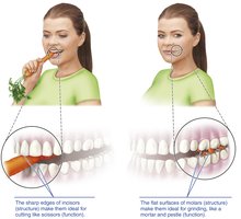
Additional info: The sharp edges of incisors are ideal for cutting, while the flat surfaces of molars are suited for grinding food, illustrating the complementarity of structure and function.
Defining Anatomy
Anatomy can be studied at various levels and with different approaches:
Gross (macroscopic) anatomy: Structures visible to the naked eye.
Regional anatomy: All structures in a specific area (e.g., head, chest).
Systemic anatomy: Study of one body system at a time (e.g., cardiovascular, nervous).
Surface anatomy: Study of internal structures as they relate to the skin surface.
Microscopic and Developmental Anatomy
Some anatomical structures are too small to be seen without magnification, and others change over time:
Microscopic anatomy: Structures too small for the naked eye.
Cytology: Study of cells.
Histology: Study of tissues.
Developmental anatomy: Structural changes throughout the lifespan (e.g., embryology).
Studying Anatomy
Mastery of anatomical terminology and practical skills is essential:
Observation and manipulation of structures.
Palpation: Feeling organs with hands.
Auscultation: Listening to body sounds (e.g., with a stethoscope).
Medical imaging: X-ray, CT, MRI, and ultrasound provide internal views without surgery.
Defining Physiology
Physiology focuses on the function of organ systems, often at the cellular or molecular level, and relies on principles of chemistry and physics (e.g., electrical currents, pressure, osmosis).
Complementarity of Structure and Function
Function always reflects structure. For example, bones support and protect due to their hard mineralized matrix, and blood flows in one direction because valves prevent backflow.
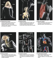
Anatomic Variability
Most human structures match textbook descriptions, but minor variations exist (e.g., nerves or vessels in different locations). Extreme variations are rare and often incompatible with life.
Sex and Gender
Sex: Biological attributes (chromosomes, hormones, reproductive anatomy).
Gender: Psychosocial construct (behaviors, expressions, identities).
Both influence health and clinical presentation.
Levels of Structural Organization
Hierarchy of Organization
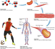
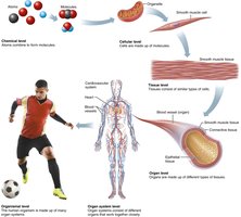
The human body is organized from simple to complex levels, each building on the previous:
Chemical level: Atoms combine to form molecules.
Cellular level: Cells are the basic units of life, composed of molecules and organelles.
Tissue level: Groups of similar cells perform common functions (epithelial, connective, muscle, nervous).
Organ level: Organs are made of two or more tissue types working together.
Organ system level: Groups of organs cooperate for a common purpose.
Organismal level: The living human being, with all systems functioning together.
Chemical Level
Atoms (e.g., H, O, C) combine to form molecules (e.g., H2O, proteins, lipids).
Molecules determine cellular structure and function (e.g., ATP stores/releases energy).
Cellular Level
Cells are the basic structural and functional units of life.
Each cell type has a unique shape and specialized function (e.g., muscle cells contract due to actin and myosin fibers).
Tissue Level
Four primary tissue types: epithelial (covering/lining), connective (support/binding), muscle (movement), nervous (communication/control).
Function depends on arrangement and composition of cells and matrix.
Organ Level
Organs are structures with two or more tissue types working together for specific functions (e.g., stomach: epithelial for secretion, muscle for mixing, connective for support, nervous for control).
Organ System Level
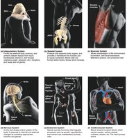
11 major systems: integumentary, skeletal, muscular, nervous, endocrine, cardiovascular, lymphatic, respiratory, digestive, urinary, reproductive.
Systems work together to support the organism.
Organismal Level
The highest level of organization: the living human being.
All organ systems function together to maintain life (e.g., respiratory and cardiovascular systems deliver oxygen to cells).
Interrelationships Among Body Systems
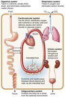
No system works in isolation; systems exchange materials and information to preserve homeostasis. For example, nutrients absorbed by the digestive system are circulated by the cardiovascular system and used by cells.
Requirements for Life
Vital Functions
Boundaries: Separation between internal and external environments (e.g., plasma membranes, skin).
Movement: Skeletal muscle enables body movement; cardiac and smooth muscle move blood, food, and other materials.
Responsiveness: Ability to sense and respond to stimuli (e.g., withdrawal reflex, changes in breathing rate).
Digestion: Breakdown of food into molecules absorbed into blood for distribution to cells.
Metabolism: Catabolism (breakdown) and anabolism (synthesis); cellular respiration produces ATP.
Excretion: Removal of metabolic and digestive wastes (e.g., urea, CO2, feces).
Reproduction: Cellular (cell division) and organismal (offspring production).
Growth: Increase in size or number of cells.
Survival Needs
Nutrients: Energy and building materials (carbohydrates, proteins, fats, vitamins, minerals).
Oxygen: Required for ATP production.
Water: Most abundant substance in the body; medium for reactions.
Normal temperature: Affects reaction rates.
Atmospheric pressure: Needed for adequate gas exchange in lungs.
Homeostasis
Definition and Importance
Homeostasis is the maintenance of stable internal conditions despite external changes. It is a state of dynamic equilibrium, with internal conditions varying within narrow limits. Homeostasis is essential for survival and proper cell function, and is mainly controlled by the nervous and endocrine systems.
Control Mechanisms
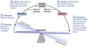
Receptor (sensor): Monitors environment and detects stimuli.
Control center: Receives input, determines set point, sends information to effector.
Effector: Provides the means for response.
Response either reduces (negative feedback) or enhances (positive feedback) the stimulus.
Negative Feedback
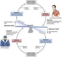
Most homeostatic controls operate via negative feedback, where the response reduces or shuts off the original stimulus, promoting stability (a self-correcting loop).
Positive Feedback
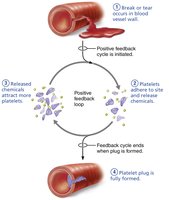
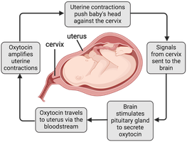
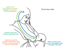
Positive feedback enhances the original stimulus, causing the variable to change in the same direction as the initial change. Examples include labor contractions (oxytocin release) and platelet plug formation during clotting.
Homeostatic Imbalance
Disturbance of homeostasis increases the risk of disease and is associated with aging. Severe imbalance may overwhelm negative feedback and allow destructive positive feedback (e.g., heart failure).
Anatomical Terminology and Body Orientation
Anatomical Position
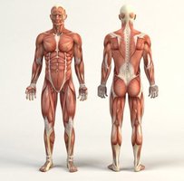
Standardized reference position: body erect, feet slightly apart, palms facing forward, thumbs outward. Right and left refer to the subject's sides, not the observer's.
Directional Terminology
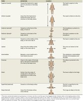
Describes one body structure in relation to another. Common pairs include:
Term | Definition | Example |
|---|---|---|
Superior (cranial) | Toward the head/upper part | The head is superior to the abdomen. |
Inferior (caudal) | Away from the head/lower part | The navel is inferior to the chin. |
Anterior (ventral) | Toward the front of the body | The breastbone is anterior to the spine. |
Posterior (dorsal) | Toward the back of the body | The heart is posterior to the breastbone. |
Medial | Toward the midline | The heart is medial to the arm. |
Lateral | Away from the midline | The arms are lateral to the chest. |
Proximal | Closer to the origin of a body part | The elbow is proximal to the wrist. |
Distal | Farther from the origin | The knee is distal to the thigh. |
Superficial | Toward the body surface | The skin is superficial to the muscles. |
Deep | Away from the body surface | The lungs are deep to the skin. |
Regional Terminology
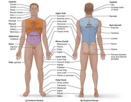
The body is divided into axial (head, neck, trunk) and appendicular (limbs) regions. Specific terms describe anterior and posterior body regions.
Body Planes
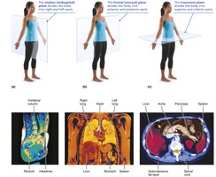
Imaginary flat surfaces used to divide the body or organs for anatomical study and imaging:
Sagittal plane: Divides body into right and left (midsagittal = midline; parasagittal = offset from midline).
Frontal (coronal) plane: Divides into anterior and posterior parts.
Transverse (horizontal) plane: Divides into superior and inferior parts.
Body Cavities and Membranes
Major Body Cavities
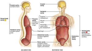
The body contains internal cavities that house and protect organs, allowing movement and expansion. Two main divisions:
Dorsal body cavity: Cranial (brain) and vertebral (spinal cord), lined by meninges.
Ventral body cavity: Thoracic (lungs, heart) and abdominopelvic (digestive, reproductive organs), separated by the diaphragm.
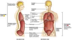
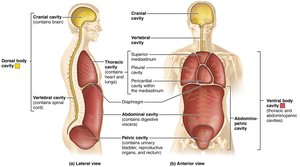
Thoracic and Abdominopelvic Cavities
Thoracic cavity: Pleural cavities (lungs), mediastinum (pericardial cavity, trachea, esophagus, thymus), pericardial cavity (heart).
Abdominopelvic cavity: Abdominal (stomach, intestines, spleen, liver) and pelvic (bladder, reproductive organs, rectum); no physical separation between the two.
Clinical Relevance: Hernias
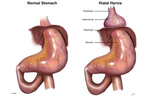
Hiatal hernia: Stomach pushes into thoracic cavity, causing acid reflux.
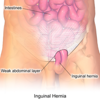
Inguinal hernia: Intestines push through the abdominal wall into the pelvic cavity.
Serous Membranes
Thin double layers lining the ventral cavity and its organs:
Parietal serosa: Lines cavity walls.
Visceral serosa: Covers organs.
Serous fluid: Lubricates and reduces friction between layers.
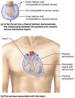
Other Serous Membranes
Pleura: Lungs (parietal lines cavity, visceral covers lungs).
Pericardium: Heart (parietal lines cavity, visceral covers heart).
Peritoneum: Abdominopelvic organs (parietal lines wall, visceral covers organs).
Inflammation can cause pain and restrict organ movement.
Abdominopelvic Quadrants and Regions
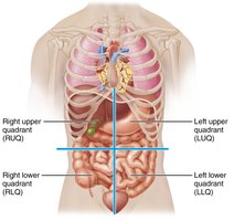
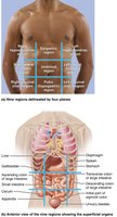
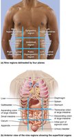
Used clinically and anatomically to localize pain or pathology:
Quadrants: Right upper (RUQ), left upper (LUQ), right lower (RLQ), left lower (LLQ).
Nine regions: Epigastric, umbilical, hypogastric (middle column); right/left hypochondriac (top sides); right/left lumbar (middle sides); right/left inguinal (bottom sides).
Clinical Relevance – Wrong Surgery Site
Errors in anatomical terminology or orientation can lead to critical mistakes, such as surgery on the wrong body part or removal of the incorrect organ lobe.
Additional Body Cavities
Oral and digestive cavities: Mouth to anus.
Nasal cavity: Respiratory passage.
Orbital cavities: Eyes.
Middle ear cavities: Hearing.
Synovial cavities: Movable joints.
