 Back
BackChapter 11: Fundamentals of the Nervous System and Nervous Tissue – Study Notes
Study Guide - Smart Notes
 Tailored notes based on your materials, expanded with key definitions, examples, and context.
Fundamentals of the Nervous System and Nervous Tissue
Overview of the Nervous System
The nervous system is the master controlling and communicating system of the body. It is responsible for monitoring internal and external stimuli, integrating sensory information, and generating motor output to effectors such as muscles and glands.
Sensory Input: Detection of changes (stimuli) inside and outside the body.
Integration: Processing and interpretation of sensory input to determine an appropriate response.
Motor Output: Activation of effector organs (muscles and glands) to produce a response.
Organization of the Nervous System
Central Nervous System (CNS): Consists of the brain and spinal cord; serves as the integration and command center.
Peripheral Nervous System (PNS): Composed of paired spinal and cranial nerves; carries messages to and from the CNS.
Functional Divisions of the PNS
Sensory (Afferent) Division: Transmits impulses from sensory receptors to the CNS.
Somatic Sensory Fibers: Carry impulses from skin, skeletal muscles, and joints.
Visceral Sensory Fibers: Transmit impulses from visceral organs.
Motor (Efferent) Division: Transmits impulses from the CNS to effector organs.
Somatic Nervous System: Voluntary control of skeletal muscles.
Autonomic Nervous System (ANS): Involuntary control of smooth muscle, cardiac muscle, and glands. Subdivided into sympathetic and parasympathetic divisions.
Histology of Nerve Tissue
Principal Cell Types
Neurons: Excitable cells that transmit electrical signals.
Supporting Cells (Neuroglia or Glial Cells): Non-excitable cells that support, protect, and insulate neurons.
Neuroglia (Supporting Cells)
Neuroglia are smaller than neurons, outnumber them, and are essential for neuron function. There are six types: four in the CNS and two in the PNS.
Astrocytes (CNS): Most abundant; support and anchor neurons, regulate the blood-brain barrier, guide neuron development, and control the chemical environment.
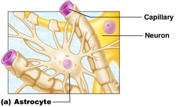
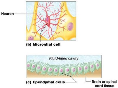
Microglia (CNS): Small, phagocytic cells that monitor neuron health and remove debris.
Ependymal Cells (CNS): Line brain and spinal cord cavities; involved in cerebrospinal fluid production and circulation.
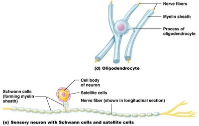
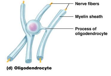
Oligodendrocytes (CNS): Form myelin sheaths around CNS nerve fibers.
Schwann Cells (PNS): Form myelin sheaths around PNS nerve fibers.
Satellite Cells (PNS): Surround neuron cell bodies in ganglia; functionally similar to astrocytes.
Neurons (Nerve Cells)
Structure of Neurons
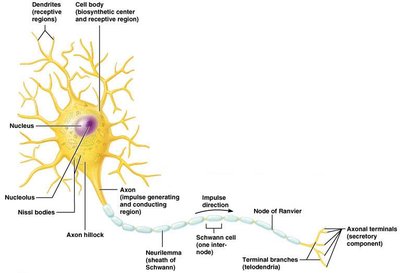
Neurons are the structural and functional units of the nervous system. They are long-lived, amitotic, and have a high metabolic rate. Each neuron consists of a cell body (soma), dendrites, and an axon.
Cell Body (Soma): Contains the nucleus, nucleolus, and organelles; major biosynthetic center; lacks centrioles (amitotic); contains Nissl bodies (rough ER).
Dendrites: Short, branched processes that receive input and convey graded potentials toward the cell body.
Axon: Single, long process that generates and transmits action potentials; ends in axon terminals (secretory region).
Myelin Sheath
The myelin sheath is a segmented, whitish, fatty covering around most long axons. It protects and insulates axons and increases the speed of nerve impulse transmission.
Formation in PNS: Schwann cells wrap around axons, forming concentric layers of membrane (myelin sheath). The outermost layer is the neurilemma.
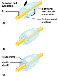
Nodes of Ranvier: Gaps between adjacent Schwann cells where axon collaterals may emerge; important for saltatory conduction.
Myelination in CNS: Oligodendrocytes form myelin sheaths; no neurilemma is present.
Gray and White Matter
White Matter: Dense collections of myelinated fibers.
Gray Matter: Mostly neuron cell bodies and unmyelinated fibers.
Classification of Neurons
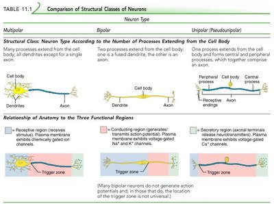
Structural Classification
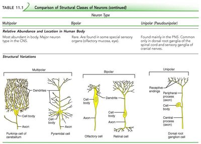
Multipolar: Three or more processes; most common in CNS.
Bipolar: Two processes (axon and dendrite); found in special sensory organs.
Unipolar (Pseudounipolar): Single process that divides into peripheral and central branches; sensory neurons in PNS.
Neuron Type | Multipolar | Bipolar | Unipolar (Pseudounipolar) |
|---|---|---|---|
Processes | Many dendrites, one axon | One dendrite, one axon | Single process splits into two branches |
Location | CNS (most abundant) | Special senses (eye, ear) | Sensory neurons in PNS |
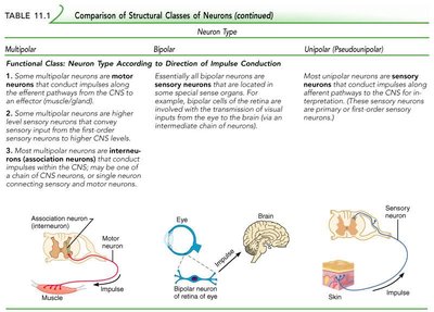
Functional Classification
Sensory (Afferent) Neurons: Transmit impulses toward the CNS.
Motor (Efferent) Neurons: Carry impulses away from the CNS to effectors.
Interneurons (Association Neurons): Connect sensory and motor neurons within the CNS.
Neurophysiology
Action Potentials
Neurons are highly excitable and communicate via action potentials—electrical impulses that travel along axons. This is the fundamental mechanism for nervous system function.
Nerve Fiber Classification
Diameter: Larger fibers conduct impulses faster.
Degree of Myelination: Myelinated fibers conduct faster than unmyelinated.
Speed of Conduction: Depends on both diameter and myelination.
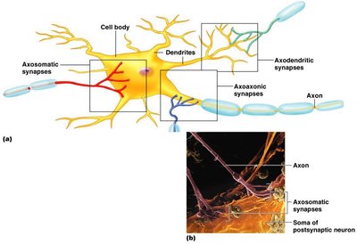
Synapses
Structure and Function
A synapse is a junction that mediates information transfer from one neuron to another or to an effector cell. The presynaptic neuron conducts impulses toward the synapse, while the postsynaptic neuron transmits impulses away from the synapse.
Types of Synapses:
Axodendritic: Between axon and dendrite.
Axosomatic: Between axon and soma (cell body).
Axoaxonic: Between axon and axon.
Electrical vs. Chemical Synapses
Electrical Synapses: Less common; involve direct cytoplasmic connections (gap junctions); allow rapid, bidirectional communication; important in CNS for arousal, attention, and memory.
Chemical Synapses: Specialized for neurotransmitter release and reception; unidirectional communication.
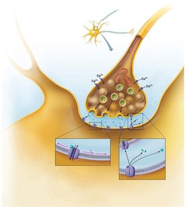
Chemical Synapse Transmission
Action potential arrives at the axon terminal of the presynaptic neuron.
Voltage-gated Ca2+ channels open, allowing Ca2+ influx.
Ca2+ triggers synaptic vesicles to release neurotransmitter by exocytosis.
Neurotransmitter diffuses across the synaptic cleft and binds to receptors on the postsynaptic membrane.
Binding opens ion channels, generating a graded potential in the postsynaptic neuron.
Neurotransmitter effects are terminated by enzymatic degradation, reuptake, or diffusion away from the synapse.
Termination of Neurotransmitter Effects
Neurotransmitters are removed from the synaptic cleft by:
Enzymatic degradation
Reuptake by astrocytes or presynaptic terminals
Diffusion away from the synapse
Additional info: The notes above provide a comprehensive overview of the structure and function of the nervous system, including the roles of different cell types, neuron structure, myelination, and synaptic transmission, as relevant to an introductory college-level anatomy and physiology course.
