 Back
BackChapter 11: Introduction to the Nervous System and Nervous Tissue – Study Notes
Study Guide - Smart Notes
 Tailored notes based on your materials, expanded with key definitions, examples, and context.
Overview of the Nervous System
General Functions
The nervous system is the primary control center for perception, voluntary movement, consciousness, learning, memory, and homeostasis. It works closely with the endocrine system to regulate vital functions such as respiratory rate, blood pressure, body temperature, sleep/wake cycles, and blood pH.
Controls voluntary and involuntary actions
Seat of consciousness and cognition
Regulates homeostasis
Anatomical Divisions
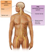
Central Nervous System (CNS): Consists of the brain and spinal cord. The brain contains billions of neurons and is protected by the skull, while the spinal cord extends from the foramen magnum to the lumbar vertebrae, enabling communication between the brain and the body below the head and neck.
Peripheral Nervous System (PNS): Includes all nerves outside the CNS. Nerves are bundles of axons, blood vessels, and connective tissue. The PNS is divided into cranial nerves (12 pairs) and spinal nerves (31 pairs).
Functional Divisions
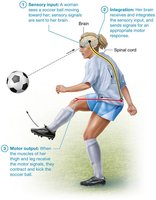
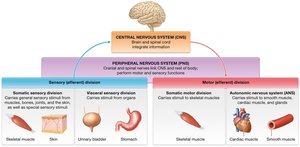
Sensory (Afferent) Division: Gathers information from internal and external environments and transmits it to the CNS.
Integrative Functions: Analyze and interpret sensory information to determine appropriate responses. Most sensory input is subconsciously disregarded.
Motor (Efferent) Division: Carries out responses via motor neurons to effectors (muscles and glands). Subdivided into:
Somatic Motor Division: Voluntary control of skeletal muscles.
Autonomic Nervous System (ANS): Involuntary control of smooth muscle, cardiac muscle, and glands.
Nervous Tissue
Neurons: Structure and Function
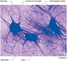
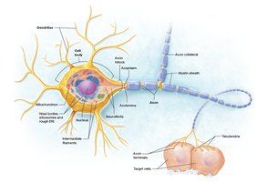
Neurons are excitable cells responsible for transmitting electrical signals (action potentials). They have three main parts:
Cell Body (Soma): Contains the nucleus, organelles, and is the site of most metabolic activity. Includes Nissl bodies (rough ER), Golgi apparatus, nucleoli, and mitochondria.
Dendrites: Short, branched processes that receive input from other neurons and transmit it toward the cell body.
Axon: A single, long process that conducts action potentials away from the cell body. Key regions include the axon hillock, axon collaterals, telodendria, and axon terminals (synaptic bulbs).
Axonal Transport
Slow Axonal Transport: Moves cytoskeletal proteins at 1–3 mm/day.
Fast Axonal Transport: Uses motor proteins and ATP to move vesicles and organelles at up to 400 mm/day (anterograde and retrograde).
Functional Regions of Neurons
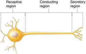
Receptive Region: Dendrites and cell body
Conducting Region: Axon
Secretory Region: Axon terminals
Neuron Classification
Structural:
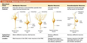
Multipolar: One axon, multiple dendrites (most common)
Bipolar: One axon, one dendrite (eye, olfactory epithelium)
Pseudounipolar: Single fused axon that splits (sensory neurons)
Functional:
Sensory (Afferent): Carry information to CNS
Interneurons: Relay information within CNS
Motor (Efferent): Carry information from CNS to effectors
Structural Class | Multipolar Neurons | Bipolar Neurons | Pseudounipolar Neurons |
|---|---|---|---|
Features | One axon, many dendrites | One axon, one dendrite | Single short process splits into two axons |
Location | CNS, motor neurons in PNS | Special sensory organs | Sensory neurons in PNS |
Neuron Groupings
CNS: Nuclei (cell bodies), tracts (axons)
PNS: Ganglia (cell bodies), nerves (axons)
Neuroglia (Neuroglial Cells)
Types and Functions
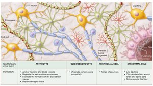
Neuroglia provide support, protection, and maintenance for neurons. They can divide and fill spaces left by dead neurons.
CNS Neuroglia:
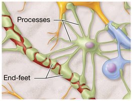
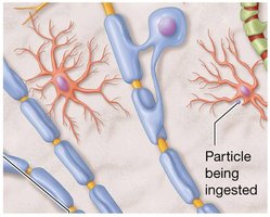
Astrocytes: Anchor neurons, regulate environment, form blood-brain barrier, repair tissue
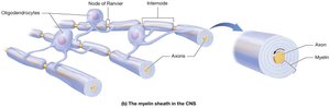
Oligodendrocytes: Myelinate axons in CNS
Microglia: Phagocytic cells, remove debris
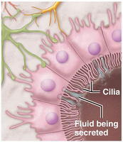
Ependymal Cells: Line CNS cavities, produce and circulate cerebrospinal fluid
PNS Neuroglia:
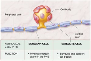
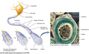
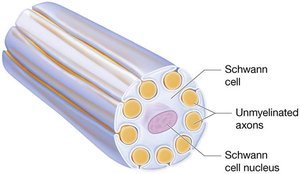
Schwann Cells: Myelinate axons in PNS
Satellite Cells: Support cell bodies in PNS
The Myelin Sheath
Structure and Function
The myelin sheath is formed by layers of plasma membrane from Schwann cells (PNS) or oligodendrocytes (CNS). It insulates axons, increasing the speed of action potential conduction.
Myelinated axons: Conduct action potentials 15–20 times faster than unmyelinated axons.
Internodes: Segments covered by myelin
Node of Ranvier: Gaps between myelinated segments
White matter: Myelinated axons
Gray matter: Cell bodies, unmyelinated dendrites and axons
Regeneration of Nervous Tissue
Regeneration in PNS
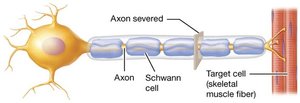
Regeneration is limited in the PNS and nearly nonexistent in the CNS. It can occur only if the cell body remains intact.
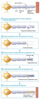
Wallerian degeneration: Axon and myelin sheath degenerate distal to injury
Growth processes form from the proximal end
Schwann cells and basal lamina form a regeneration tube
New axon grows toward target cell and reconnects
Electrophysiology of Neurons
Resting Membrane Potential
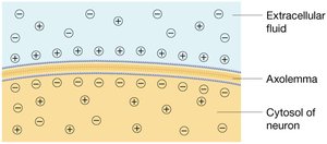
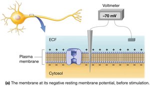
Neurons maintain a resting membrane potential (RMP) of approximately –70 mV, with a thin layer of negative ions inside and positive ions outside the membrane.
Voltage: Electrical gradient across the membrane
Membrane potential: Source of potential energy for the cell
Ion Channels and Gradients
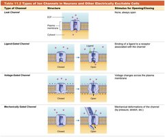
Leak Channels: Always open, allow ions to flow down gradients
Gated Channels: Open in response to specific stimuli
Ligand-gated: Open when a chemical binds
Voltage-gated: Open with voltage changes
Mechanically-gated: Open with mechanical stimulation
Changes in Membrane Potential
Depolarization: Membrane potential becomes less negative (e.g., –70 mV to –60 mV)
Repolarization: Return to resting potential
Hyperpolarization: Membrane potential becomes more negative (e.g., –70 mV to –80 mV)
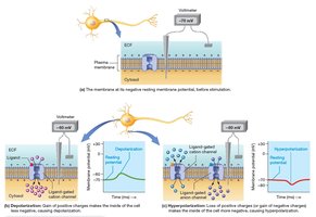
Local Potentials
Small, graded changes in membrane potential
Reversible and decremental (decrease over distance)
Trigger action potentials if threshold is reached
Action Potentials
Phases and Mechanisms
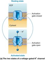
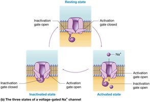
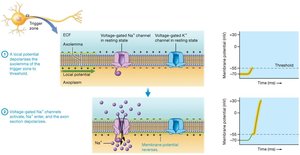
Action potentials are rapid, uniform changes in membrane potential that travel along axons. They involve voltage-gated sodium and potassium channels.
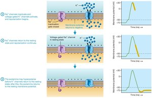
Depolarization: Sodium channels open, Na+ enters
Repolarization: Sodium channels inactivate, potassium channels open, K+ exits
Hyperpolarization: Potassium channels remain open briefly, membrane potential becomes more negative than RMP
Refractory Periods
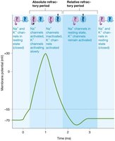
Absolute Refractory Period: No new action potential can be generated
Relative Refractory Period: Only a strong stimulus can trigger another action potential
Propagation of Action Potentials
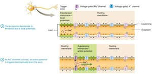
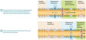
Action potentials are self-propagating and travel in one direction (from trigger zone to axon terminals)
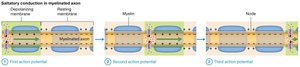
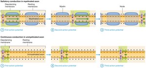
Saltatory Conduction: In myelinated axons, action potentials jump between nodes of Ranvier, increasing speed
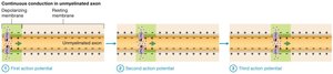
Continuous Conduction: In unmyelinated axons, action potentials propagate along every segment, slowing conduction
Neuronal Synapses
Types of Synapses
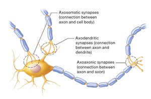
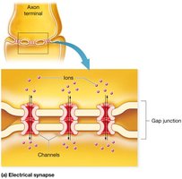
Electrical Synapses: Direct flow of ions via gap junctions; bidirectional and nearly instantaneous; found in brain regions for automatic behaviors and in cardiac/smooth muscle
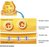
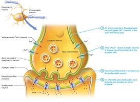
Chemical Synapses: Use neurotransmitters to transmit signals across a synaptic cleft; unidirectional and allow for variable signal strength
Postsynaptic Potentials
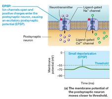
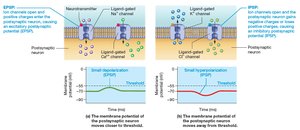
Excitatory Postsynaptic Potential (EPSP): Small depolarization, brings membrane closer to threshold (Na+ or Ca2+ influx)
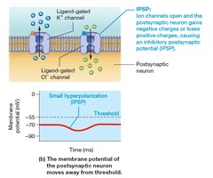
Inhibitory Postsynaptic Potential (IPSP): Small hyperpolarization, moves membrane away from threshold (K+ efflux or Cl– influx)
Neural Integration and Summation
Neural Integration: Postsynaptic neuron integrates all incoming EPSPs and IPSPs
Summation: Temporal (rapid, repeated signals from one neuron) and spatial (simultaneous signals from multiple neurons)
Action potential is generated only if threshold is reached at the trigger zone
Termination of Synaptic Transmission
Neurotransmitters are removed by diffusion, enzymatic degradation, or reuptake into the presynaptic neuron
Neurotransmitters
General Features
Made in the cell body or axon terminal, packaged into vesicles
Released into the synaptic cleft, bind to receptors on the postsynaptic membrane
Effects are rapidly terminated by removal or degradation
Example: Acetylcholine (ACh)
Widely used in the nervous system (neuromuscular junction, brain, spinal cord, autonomic nervous system)
Mostly excitatory, but can be inhibitory in the PNS
Degraded by acetylcholinesterase (AChE)
Functional Groups of Neurons
Neuronal Pools and Circuits
Neuronal Pools: Groups of interneurons that process specific types of information
Diverging Circuits: One neuron communicates with multiple targets (e.g., sensory information distribution)
Converging Circuits: Multiple neurons converge on a single postsynaptic neuron (e.g., motor control)
Inhibitory circuits and synaptic fatigue help stabilize neural activity
Additional info: This guide covers the foundational concepts of nervous tissue, neuron structure and function, neuroglia, myelination, electrophysiology, synaptic transmission, neurotransmitters, and neural circuits, as outlined in a typical Anatomy & Physiology curriculum.
