 Back
BackChapter 12: Muscles – Structure, Function, and Physiology
Study Guide - Smart Notes
 Tailored notes based on your materials, expanded with key definitions, examples, and context.
Muscle Overview
Types of Muscle Tissue
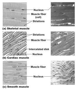
Muscle tissue is specialized for contraction and is essential for movement, posture, and various physiological functions. There are three main types of muscle tissue in the human body:
Skeletal Muscle: Voluntary, striated muscle attached to bones for movement and posture.
Cardiac Muscle: Involuntary, striated muscle found only in the heart, responsible for pumping blood.
Smooth Muscle: Involuntary, non-striated muscle found in walls of internal organs, responsible for movements such as peristalsis and vasoconstriction.
Organization of Skeletal Muscle
Hierarchical Structure
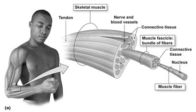
Skeletal muscle is organized into a hierarchy of structures, from the whole muscle down to the molecular level:
Muscle: Surrounded by connective tissue, containing bundles of fascicles.
Fascicle: Bundle of muscle fibers (cells).
Muscle Fiber: Multinucleated cell containing myofibrils.
Myofibril: Composed of repeating units called sarcomeres, the contractile units of muscle.
Sarcomere: Contains thick (myosin) and thin (actin) filaments, along with regulatory and structural proteins.
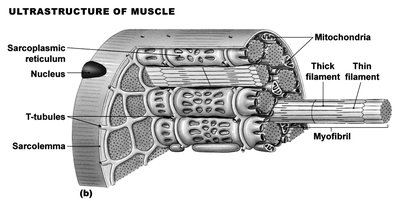
Muscle Fiber Structure
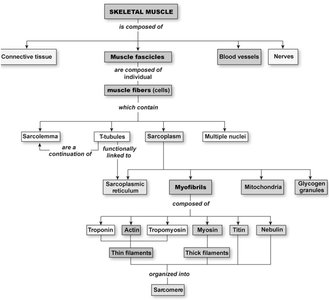
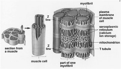
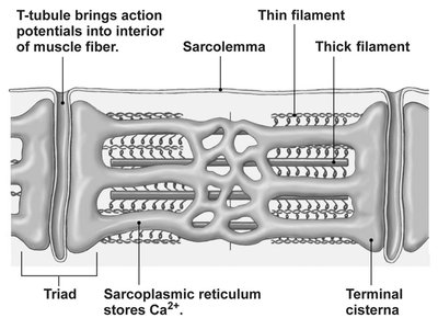
Sarcolemma: Plasma membrane of the muscle fiber.
T-tubules: Invaginations of the sarcolemma that transmit action potentials into the fiber.
Sarcoplasmic Reticulum (SR): Specialized endoplasmic reticulum that stores and releases Ca2+ for contraction.
Mitochondria: Provide ATP for contraction.
Sarcomere Structure and Proteins
Sarcomere Organization
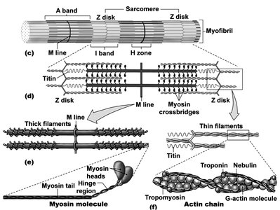
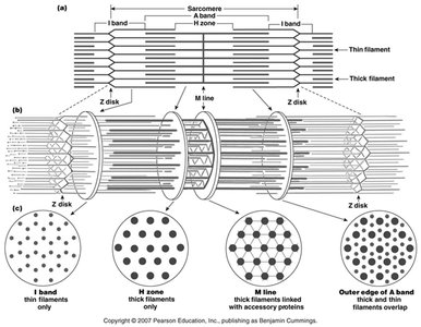
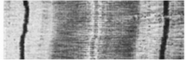
The sarcomere is the functional unit of muscle contraction, defined by the region between two Z-disks. It contains:
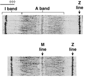
Z disk: Anchors thin filaments and defines sarcomere boundaries.
I band: Contains only thin filaments (actin).
A band: Contains the entire length of thick filaments (myosin), including regions of overlap with thin filaments.
H zone: Central region of A band with only thick filaments.
M line: Center of the sarcomere, where thick filaments are linked.

Key Sarcomeric Proteins
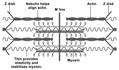
Actin: Forms thin filaments; provides binding sites for myosin.
Myosin: Motor protein forming thick filaments; interacts with actin for contraction.
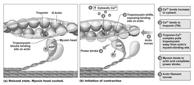
Tropomyosin: Blocks myosin-binding sites on actin in resting muscle.
Troponin: Binds Ca2+ and moves tropomyosin to expose binding sites.
Titin: Provides elasticity and stabilizes myosin.
Nebulin: Helps align actin filaments.
Sliding Filament Theory of Muscle Contraction
Mechanism of Contraction
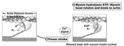
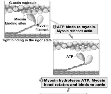
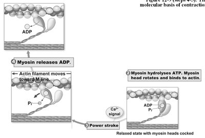
Muscle contraction occurs through the sliding of thin filaments past thick filaments, shortening the sarcomere without changing the length of the filaments themselves. This process is powered by ATP hydrolysis and regulated by Ca2+ binding to troponin.
ATP binds to myosin, causing it to release actin.
Myosin hydrolyzes ATP, cocks its head, and binds to a new position on actin.
Ca2+ binds to troponin, shifting tropomyosin and exposing myosin-binding sites on actin.
Myosin executes a power stroke, pulling actin toward the M line.
ADP and Pi are released; myosin remains bound until another ATP binds.
Excitation-Contraction Coupling
Sequence of Events
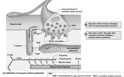
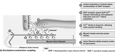
Excitation-contraction coupling links the action potential in the muscle fiber to the initiation of contraction:
Action potential arrives at the neuromuscular junction, releasing acetylcholine (ACh).
ACh binds to nicotinic receptors, causing Na+ influx and depolarization of the sarcolemma.
Action potential travels down T-tubules, activating DHP receptors.
DHP receptors mechanically open RyR channels on the SR, releasing Ca2+ into the sarcoplasm.
Ca2+ binds to troponin, initiating contraction.
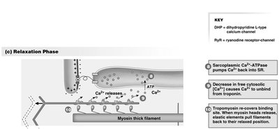
Relaxation occurs when Ca2+ is pumped back into the SR by Ca2+-ATPase.
Muscle Metabolism and Fatigue
Sources of ATP
Aerobic Respiration: Uses oxygen, glucose, and fatty acids to produce 30–32 ATP per glucose.
Anaerobic Metabolism: Fast, produces 2 ATP per glucose (lactic acid fermentation).
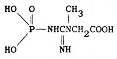
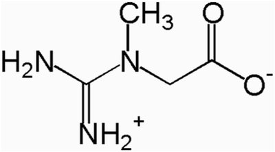
Phosphocreatine: Provides a rapid but short-term buffer for ATP (2–7 seconds).
Muscle Fatigue
Central Fatigue: Originates in the CNS; associated with the perception of tiredness.
Peripheral Fatigue: Due to changes at the neuromuscular junction or within the muscle fiber (e.g., glycogen depletion, high Pi, K+ imbalance, ACh depletion).
Muscle Fiber Types
Classification by Contraction Speed and Metabolism
Fast-Twitch Glycolytic Fibers: Rapid contraction, fatigue quickly, rely on anaerobic metabolism.
Fast-Twitch Oxidative Fibers: Intermediate speed, use both aerobic and anaerobic metabolism.
Slow-Twitch Oxidative Fibers: Slow contraction, resistant to fatigue, high in mitochondria and myoglobin, suited for endurance.
Control of Muscle Force
Motor Units and Recruitment
Motor Unit: A single motor neuron and all the muscle fibers it innervates; all fibers contract together when the neuron fires.
Recruitment: Increasing the number of active motor units increases muscle force; slow-twitch fibers are recruited first, followed by fast-twitch as needed.
Summation and Tetanus
Summation: Increased frequency of stimulation leads to greater force as contractions build on each other.
Tetanus: Sustained maximal contraction when stimuli are so frequent that relaxation does not occur.
Muscle Mechanics
Isotonic vs. Isometric Contractions
Isotonic Contraction: Muscle changes length (shortens or lengthens) while tension remains constant; produces movement.
Isometric Contraction: Muscle develops tension without changing length; stabilizes joints and maintains posture.
Levers and Body Movement
Bones act as levers and joints as fulcrums to amplify force or speed of movement.
Muscle origin is proximal, insertion is distal; arrangement affects mechanical advantage.
Smooth and Cardiac Muscle
Smooth Muscle
Found in walls of hollow organs; contracts slowly and can sustain contractions with little fatigue.
Cells are spindle-shaped, have a single nucleus, and lack striations.
Contraction can be initiated by electrical, chemical, or mechanical signals; Ca2+ binds to calmodulin (not troponin).
Cardiac Muscle
Found only in the heart; striated like skeletal muscle but involuntary.
Cells are branched, have a single nucleus, and are connected by intercalated discs (containing gap junctions and desmosomes).
Can generate action potentials spontaneously (pacemaker cells); regulated by autonomic nervous system and hormones.
Summary Table: Comparison of Muscle Types
Feature | Skeletal Muscle | Cardiac Muscle | Smooth Muscle |
|---|---|---|---|
Striations | Yes | Yes | No |
Control | Voluntary | Involuntary | Involuntary |
Cell Shape | Long, cylindrical, multinucleated | Branched, single nucleus | Spindle-shaped, single nucleus |
Location | Attached to bones | Heart | Walls of hollow organs |
Special Features | Motor units, rapid contraction | Intercalated discs, pacemaker cells | Gap junctions, slow sustained contraction |
