 Back
BackChapter 12: Nervous Tissue – Structured Study Notes for Anatomy & Physiology
Study Guide - Smart Notes
 Tailored notes based on your materials, expanded with key definitions, examples, and context.
Nervous Tissue
An Introduction to the Nervous System
The nervous system is a complex network responsible for receiving, processing, and responding to internal and external stimuli. It consists of the brain, spinal cord, sensory receptors, and nerves that connect to other body systems.
Functions:
Receives information from internal and external stimuli
Processes information and initiates responses
Cell Types:
Neurons: Specialized for intercellular communication
Neuroglia (glial cells): Support, protect, and preserve nervous tissue structure
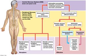
Divisions of the Nervous System
The nervous system is divided anatomically and functionally into the central and peripheral nervous systems.
Central Nervous System (CNS):
Includes brain and spinal cord
Integrates, processes, and coordinates sensory information and motor commands
Responsible for higher functions: intelligence, memory, learning, emotion
Peripheral Nervous System (PNS):
All nervous tissue outside CNS
Delivers sensory information to CNS and carries motor commands to peripheral tissues
Nerves: Bundles of axons with connective tissues and blood vessels
Cranial nerves connect to brain; spinal nerves connect to spinal cord
Functional Divisions of PNS:
Afferent Division: Carries sensory information from receptors to CNS
Efferent Division: Carries motor commands from CNS to effectors (muscles, glands, adipose tissue)
Somatic Nervous System (SNS): Controls skeletal muscle contractions (voluntary and involuntary)
Autonomic Nervous System (ANS): Controls smooth muscle, cardiac muscle, adipose tissue, glands (involuntary); includes sympathetic and parasympathetic divisions
Enteric Nervous System (ENS): Neurons in digestive tract walls, coordinates local visceral reflexes
Neurons
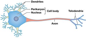
Structure and Function of Neurons
Neurons are the basic functional units of the nervous system, specialized for communication, information processing, and control.
Cell Body (Soma): Contains nucleus, perikaryon (cytoplasm), neurofilaments, neurotubules, and neurofibrils for structural support
Dendrites: Highly branched processes that receive information from other neurons
Axon: Single, long process that propagates electrical signals (action potentials); includes axoplasm, axolemma, initial segment, axon hillock, collaterals, telodendria, and axon terminals
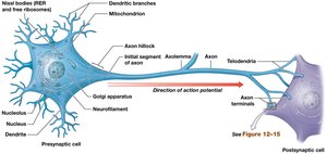
Axonal Transport
Anterograde: Movement from cell body to axon terminals
Retrograde: Movement from axon terminals to cell body (e.g., rabies virus infection)
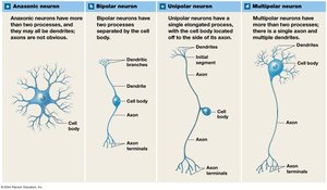
Classification of Neurons
Structural Classification:
Anaxonic: Many dendrites, no obvious axon; found in brain and special sense organs
Bipolar: One dendrite, one axon; rare, found in special sense organs
Unipolar: Axon and dendrites continuous, soma off to side; most sensory neurons in PNS
Multipolar: One long axon, two or more dendrites; common in CNS and all PNS motor neurons
Functional Classification:
Sensory (Afferent) Neurons: Carry information from receptors to CNS
Motor (Efferent) Neurons: Carry instructions from CNS to effectors
Interneurons: Integrate sensory information and coordinate motor commands; involved in higher functions
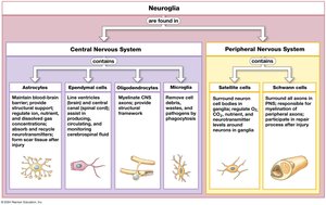
Neuroglia
Types and Functions of Neuroglia
Neuroglia are supporting cells that protect and maintain neurons, making up half the volume of the nervous system.
CNS Neuroglia:
Astrocytes: Maintain blood-brain barrier, repair tissue, guide development, regulate environment
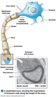
Oligodendrocytes: Form myelin sheath, increase speed of action potentials
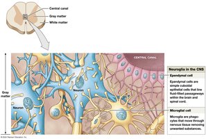
Ependymal Cells: Line ventricles and central canal, produce and monitor cerebrospinal fluid
Microglia: Phagocytic cells that clean up debris and pathogens
PNS Neuroglia:
Satellite Cells: Surround neuron cell bodies in ganglia, regulate interstitial fluid
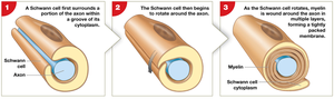
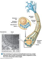
Schwann Cells: Form myelin sheath or folds around axons, aid in regeneration
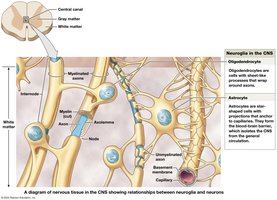
Myelination and Peripheral Nerve Regeneration
Myelin: Lipid insulation that increases speed of action potentials
Internodes: Myelinated segments
Nodes of Ranvier: Gaps in myelin sheath
White Matter: Regions with myelinated axons
Gray Matter: Regions with unmyelinated axons, cell bodies, dendrites
Membrane Potential
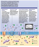
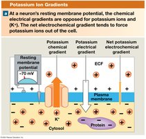
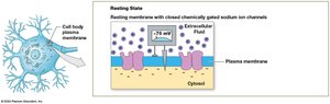
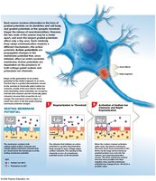
Resting Membrane Potential
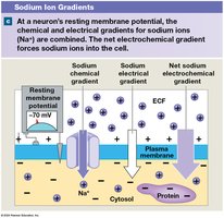
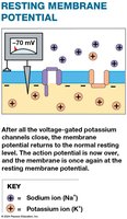
The resting membrane potential is the voltage difference across the cell membrane of an unstimulated neuron, typically around -70 mV.
Ion Distribution:
ECF: High Na+ and Cl-
Cytosol: High K+ and negatively charged proteins
Selective Permeability: More K+ leaks than Na+; proteins cannot cross membrane
Sodium-Potassium Pump: Maintains gradient by moving 3 Na+ out and 2 K+ in per ATP
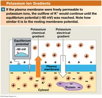
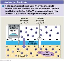
Electrochemical Gradients and Equilibrium Potential
Electrochemical Gradient: Sum of chemical and electrical forces acting on an ion
Equilibrium Potential: Membrane potential at which there is no net movement of a particular ion
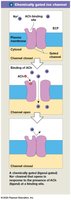
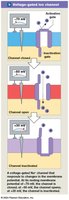
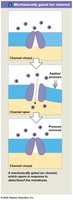
Membrane Channels
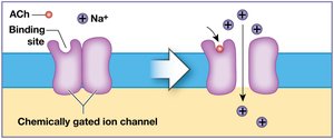
Gated Ion Channels: Open/close in response to stimuli; types include chemically gated, voltage-gated, and mechanically gated
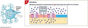
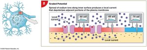
Graded Potentials
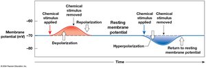
Definition: Temporary, localized changes in membrane potential
Depolarization: Shift toward less negative potential (Na+ influx)
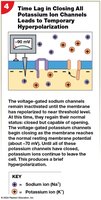
Hyperpolarization: Shift toward more negative potential (K+ efflux)
Repolarization: Return to resting potential
Action Potential
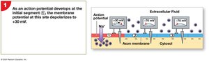
Generation and Propagation of Action Potentials
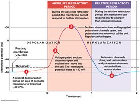
An action potential is a large depolarization that propagates along the axon, following the all-or-none principle.
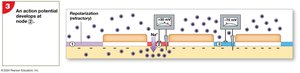
Steps:
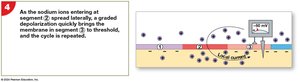
Depolarization to threshold
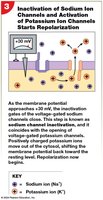
Rapid depolarization (Na+ influx)
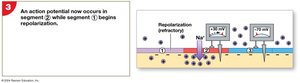
Repolarization (K+ efflux)
Hyperpolarization (brief, due to slow K+ channel closure)
Refractory Period:
Absolute: No response possible
Relative: Larger stimulus required
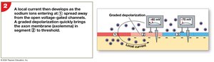
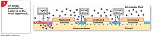
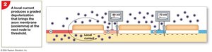
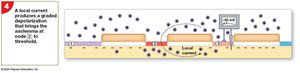
Propagation Types
Continuous Propagation: Unmyelinated axons; action potential moves stepwise
Saltatory Propagation: Myelinated axons; action potential jumps from node to node, faster and more energy-efficient
Axon Types and Propagation Speed
Type A: Myelinated, large diameter, rapid transmission (sensory info, motor impulses)
Type B: Myelinated, medium diameter, intermediate speed
Type C: Unmyelinated, small diameter, slow transmission (sensory info from skin, motor instructions to smooth/cardiac muscle and glands)
Synapses
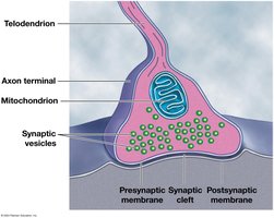
Structure and Function of Synapses
A synapse is a specialized site where a neuron communicates with another cell, either electrically or chemically.
Electrical Synapses: Direct physical contact, rapid transmission
Chemical Synapses: Use neurotransmitters to transmit signals across a synaptic cleft
Types: Axoaxonic, axosomatic, axodendritic, neuromuscular, neuroglandular
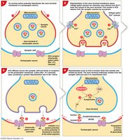
Cholinergic Synapses
Release acetylcholine (ACh)
Events: Action potential arrives, Ca2+ influx triggers ACh release, ACh binds to postsynaptic receptors, ACh broken down by acetylcholinesterase
Neurotransmitters and Neuromodulators
Types and Effects
Excitatory: Cause depolarization, promote action potentials
Inhibitory: Cause hyperpolarization, suppress action potentials
Major Classes:
Acetylcholine: Excitatory, CNS and PNS
Biogenic Amines: Norepinephrine (excitatory), dopamine (excitatory/inhibitory), serotonin (affects mood)
Amino Acids: Glutamate (excitatory), GABA (inhibitory)
Neuropeptides: Opioids (pain relief), enkephalins, endorphins, dynorphins
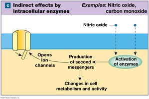
Dissolved Gases: Nitric oxide, carbon monoxide
Mechanisms of Action
Ionotropic (Direct): Open/close ion channels directly (e.g., ACh, glutamate)
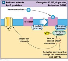
Metabotropic (Indirect): Bind to G protein-coupled receptors, activate second messengers (e.g., NE, dopamine, serotonin, GABA)
Intracellular Enzyme Activation: Lipid-soluble gases enter cell and activate enzymes (e.g., NO, CO)
Information Processing in Nervous Tissue
Postsynaptic Potentials and Summation
Excitatory Postsynaptic Potential (EPSP): Graded depolarization
Inhibitory Postsynaptic Potential (IPSP): Graded hyperpolarization
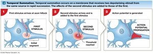
Summation:
Temporal: Rapid, repeated stimuli at a single synapse
Spatial: Simultaneous stimuli at multiple synapses
Facilitation and Presynaptic Regulation
Facilitation: Brings membrane potential closer to threshold
Presynaptic Inhibition: Decreases neurotransmitter release
Presynaptic Facilitation: Increases neurotransmitter release
Rate of Action Potential Generation
Strength of response is proportional to frequency of stimulation
Maximum rate reached when relative refractory period is eliminated
Summary
Information is relayed as action potentials
Neurotransmitters and neuromodulators can have excitatory or inhibitory effects
Neurons may be facilitated or inhibited by chemicals
Response of postsynaptic neuron can be altered by neuromodulators, activity at other synapses, or modification of neurotransmitter release
Key Equations
Nernst Equation for Equilibrium Potential
The equilibrium potential for an ion can be calculated using the Nernst equation:
For potassium (K+):
For sodium (Na+):
Summary Table: Types of Neuroglia
Neuroglia Type | Location | Main Function |
|---|---|---|
Astrocytes | CNS | Maintain BBB, repair, guide development, regulate environment |
Oligodendrocytes | CNS | Form myelin sheath |
Ependymal Cells | CNS | Produce/monitor CSF |
Microglia | CNS | Phagocytosis |
Satellite Cells | PNS | Regulate interstitial fluid |
Schwann Cells | PNS | Myelinate axons, aid regeneration |
Summary Table: Structural Classification of Neurons
Type | Structure | Location |
|---|---|---|
Anaxonic | Many dendrites, no obvious axon | Brain, special sense organs |
Bipolar | One dendrite, one axon | Special sense organs |
Unipolar | Axon and dendrites continuous, soma off to side | Sensory neurons in PNS |
Multipolar | One long axon, two or more dendrites | CNS, all PNS motor neurons |
