 Back
Chapter 12: The Brain – Structure, Function, and Protection
Study Guide - Smart Notes
 Tailored notes based on your materials, expanded with key definitions, examples, and context.
Gross Anatomy and Organization of the Adult Brain
Main Regions of the Brain
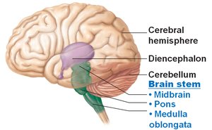
The adult brain is divided into four major regions, each with distinct anatomical and functional characteristics. Understanding these regions is essential for grasping the complexity of the central nervous system (CNS).
Cerebral Hemispheres: The largest part of the brain, responsible for higher mental functions.
Diencephalon: Contains structures such as the thalamus and hypothalamus, crucial for sensory and autonomic integration.
Brain Stem: Includes the midbrain, pons, and medulla oblongata; controls vital life functions.
Cerebellum: Coordinates movement and balance.
Structural Organization of the CNS
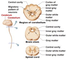
The CNS is organized into regions of gray and white matter. The arrangement of these tissues varies between the cerebrum, cerebellum, brain stem, and spinal cord.
Gray Matter: Contains neuron cell bodies; forms the cortex and nuclei.
White Matter: Composed of myelinated axons; responsible for communication between regions.
Ventricles of the Brain
Structure and Function
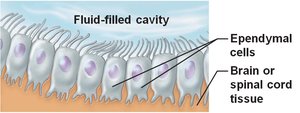
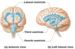
The brain contains interconnected cavities called ventricles, which are filled with cerebrospinal fluid (CSF) and lined by ependymal cells. These structures help protect and nourish the brain.
Lateral Ventricles: Two C-shaped cavities in the cerebral hemispheres.
Third Ventricle: Located in the diencephalon.
Fourth Ventricle: Found in the hindbrain, connected to the central canal of the spinal cord.
Ependymal Cells: Specialized glial cells that line the ventricles and help circulate CSF.
Cerebral Hemispheres
Surface Markings and Lobes
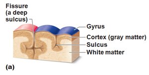
The surface of the cerebral hemispheres is highly folded, increasing surface area for cortical neurons. Key features include:
Gyri: Elevated ridges.
Sulci: Shallow grooves.
Fissures: Deep grooves separating major brain regions.
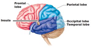
Lobes of the Brain: Frontal, parietal, temporal, occipital, and insula.
Major Sulci and Fissures
Central Sulcus: Separates frontal and parietal lobes.
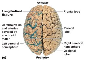
Longitudinal Fissure: Divides the two cerebral hemispheres.
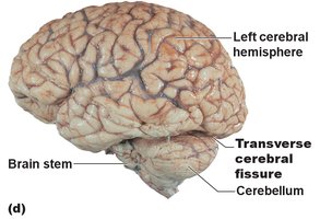
Transverse Cerebral Fissure: Separates the cerebrum from the cerebellum.
Internal Organization
The cerebral hemispheres consist of three main regions:
Cortex: Outer layer of gray matter.
White Matter: Inner layer containing myelinated fibers.
Basal Nuclei: Islands of gray matter within the white matter.
Cerebral Cortex: Functional Areas
Overview
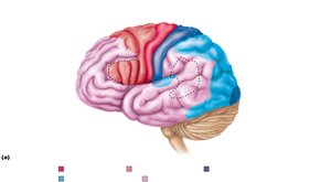
The cerebral cortex is the site of conscious mind, responsible for awareness, sensory perception, voluntary motor initiation, communication, memory storage, and understanding. Each hemisphere controls the opposite side of the body (contralateral control), and there is lateralization of function.
Motor Areas: Control voluntary movement.
Sensory Areas: Provide conscious awareness of sensation.
Association Areas: Integrate diverse information for complex functions.
Lateralization of Cortical Function
Left Hemisphere: Dominant for language, math, and logic (in 90% of people).
Right Hemisphere: Responsible for insight, visual-spatial skills, intuition, and artistic abilities.
Cerebral White Matter and Basal Nuclei
White Matter Tracts
Cerebral white matter consists of myelinated fibers that facilitate communication within the brain:
Commissural Fibers: Connect gray matter of the two hemispheres (e.g., corpus callosum).
Association Fibers: Connect different parts of the same hemisphere.
Projection Fibers: Connect the hemispheres with lower brain or spinal cord.
Diencephalon
Main Structures
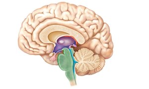
The diencephalon is composed of three paired structures:
Thalamus: Gateway to the cerebral cortex; sorts, edits, and relays sensory information.
Hypothalamus: Autonomic control center; regulates body temperature, hunger, thirst, sleep, and endocrine functions.
Epithalamus: Contains the pineal gland, which secretes melatonin to regulate sleep-wake cycles.
Brain Stem
Components and Functions
The brain stem consists of the midbrain, pons, and medulla oblongata. It is responsible for automatic behaviors necessary for survival and is the origin of most cranial nerves.
Midbrain: Contains reflex centers for vision and hearing; produces dopamine.
Pons: Connects higher brain centers to the spinal cord and relays information between the motor cortex and cerebellum.
Medulla Oblongata: Autonomic reflex center; regulates cardiovascular and respiratory functions.
Cerebellum
Structure and Function
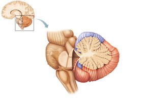
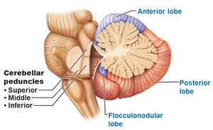
The cerebellum coordinates voluntary movements and maintains posture and balance. It consists of two hemispheres connected by the vermis, each with three lobes: anterior, posterior, and flocculonodular. The arbor vitae is the distinctive tree-like pattern of white matter.
Cerebellar Peduncles: Fiber tracts that connect the cerebellum to the brain stem.
Protection of the Brain
Protective Structures
The brain is protected by several physical and chemical barriers:
Bone: The skull encases the brain.
Meninges: Three connective tissue membranes (dura mater, arachnoid mater, pia mater) cover and protect the CNS.
Cerebrospinal Fluid (CSF): Provides a cushion and helps nourish the brain.
Blood-Brain Barrier: Regulates the passage of substances from the blood into the brain.
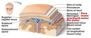
Meninges
Dura Mater: Tough, outermost layer; forms dural sinuses.
Arachnoid Mater: Middle layer with web-like extensions; contains arachnoid villi for CSF reabsorption.
Pia Mater: Delicate, innermost layer; adheres closely to the brain surface.
Cerebrospinal Fluid (CSF)
CSF is a clear, colorless fluid that surrounds the brain and spinal cord. It is produced by the choroid plexuses and serves several functions:
Provides buoyancy and protection for the CNS.
Nourishes brain tissue and removes waste.
Carries chemical signals throughout the CNS.
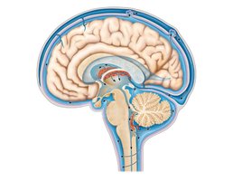
Choroid Plexuses
Choroid plexuses are clusters of capillaries surrounded by pia mater and ependymal cells. They produce CSF and help maintain its composition.
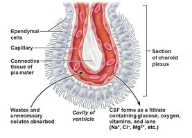
Blood-Brain Barrier
The blood-brain barrier is a selective barrier formed by endothelial cells, a basal lamina, and astrocyte end-feet. It protects the brain from harmful substances while allowing essential nutrients to pass through.
Allows nutrients and fat-soluble substances (e.g., alcohol, nicotine) to pass.
Absent in some areas (e.g., vomiting center, hypothalamus) where blood composition must be monitored.
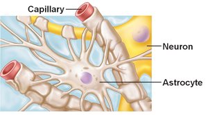
Conclusion
This chapter provides an overview of the gross anatomy and physiology of the adult brain, higher mental functions, and the protective mechanisms that maintain CNS integrity. Mastery of these concepts is foundational for understanding the central nervous system's role in human physiology.
