 Back
BackChapter 14: The Brain and Cranial Nerves – Structured Study Notes
Study Guide - Smart Notes
 Tailored notes based on your materials, expanded with key definitions, examples, and context.
The Brain and Cranial Nerves
Introduction to the Brain
The adult human brain is the central organ of the nervous system, containing nearly 97% of the body's nervous tissue. It weighs approximately 1.4 kg and has a typical volume of 1200 mL, though normal ranges vary. Despite size differences, there is no correlation between brain size and intelligence.
Key Point: The brain is divided into several major regions, each with distinct functions.
Key Point: The brain is protected by physical and biochemical mechanisms.
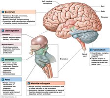
Major Regions of the Brain
The brain is organized into four primary regions: the cerebrum, cerebellum, diencephalon, and brainstem. Each region is responsible for specific functions and processes.
Cerebrum: Largest part; controls sensations, complex movement, and higher mental functions (conscious thought, intellect, memory). Divided into left and right hemispheres. The cerebral cortex is the highly folded superficial layer of gray matter.
Cerebellum: Second-largest; coordinates ongoing body movements and balance. Covered by gray matter (cerebellar cortex).
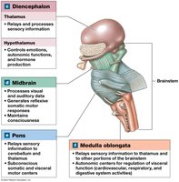
Diencephalon: Contains the thalamus (sensory relay) and hypothalamus (emotions, autonomic functions, hormone production).
Brainstem: Processes and relays information between the spinal cord and higher brain regions. Includes the midbrain, pons, and medulla oblongata.
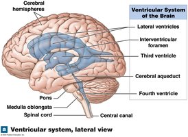
Ventricular System of the Brain
The brain contains internal chambers called ventricles, lined with ependymal cells. These ventricles are filled with cerebrospinal fluid (CSF) and are essential for brain protection and support.
Key Point: The ventricular system consists of the lateral, third, and fourth ventricles, connected by the cerebral aqueduct and central canal.
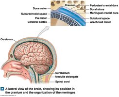
Brain Protection and Support
Physical and Biochemical Protection
The brain is protected by the bones of the skull, cranial meninges, and cerebrospinal fluid. Biochemical protection is provided by the blood-brain barrier.
Cranial Meninges: Three layers – dura mater (outer), arachnoid mater (middle), and pia mater (inner). The subarachnoid space lies between the arachnoid and pia mater.
Cerebrospinal Fluid (CSF): Surrounds all exposed surfaces of the CNS, supports and cushions the brain, and transports nutrients and wastes.
Blood-Brain Barrier (BBB): Isolates the CNS from general circulation, allowing selective transport of substances.
Formation and Circulation of Cerebrospinal Fluid
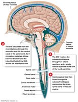
CSF is produced by the choroid plexus in each ventricle. Specialized ependymal cells secrete and regulate CSF composition, removing wastes and adjusting its chemical makeup.
Key Point: CSF circulates through the ventricles, central canal, and subarachnoid space, providing buoyancy and protection.
Blood-Brain and Blood-CSF Barriers
The BBB is regulated by astrocytes and restricts passage of most substances except lipid-soluble compounds. The blood-CSF barrier, formed by tight junctions between ependymal cells, limits transfer of substances to CSF, allowing its composition to differ from blood.
Example: Oxygen and carbon dioxide can diffuse freely, while glucose is actively transported.
The Brainstem
The Medulla Oblongata
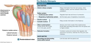
The medulla oblongata is the most inferior part of the brainstem, connecting the brain to the spinal cord. It contains nuclei for autonomic reflexes, cranial nerves, and relay stations for sensory and motor pathways.
Reflex Centers: Regulate autonomic functions such as heart rate, blood pressure, and breathing.
Sensory and Motor Nuclei: Associated with five cranial nerves.
The Pons
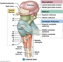
The pons contains nuclei for cranial nerves, centers for respiratory control (apneustic and pneumotaxic), and tracts that relay information to and from the cerebellum. It links the cerebellum with other brain regions and the spinal cord.
The Midbrain
The midbrain processes visual and auditory information and contains important nuclei:
Red Nucleus: Issues subconscious motor commands for upper limbs.
Substantia Nigra: Produces dopamine and inhibits basal nuclei activity; affected in Parkinson's disease.
Cerebral Peduncles: Contain descending motor fibers.
The Cerebellum
Anatomy and Functions
The cerebellum is the center for automatic processing and coordination of muscular activity. It contains the cerebellar cortex (gray matter), anterior and posterior lobes, and the arbor vitae (internal white matter).
Key Functions: Adjusts postural muscles, programs and fine-tunes movements, refines learned movement patterns, and coordinates motor commands with proprioceptive information.
Disorders: Ataxia – disturbance in muscular coordination, often caused by trauma, stroke, or alcohol intoxication.
The Diencephalon
Anatomy and Functions
The diencephalon integrates sensory information with motor commands at the subconscious level. It contains the epithalamus, thalamus, and hypothalamus.
Epithalamus: Roof of the diencephalon; contains the pineal gland, which secretes melatonin.
Thalamus: Lateral walls; relays and filters sensory information to the cerebral cortex.
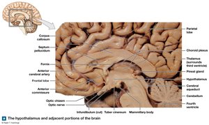
Hypothalamus: Floor; regulates emotions, autonomic functions, and links nervous and endocrine systems. Contains the infundibulum (connects to pituitary gland) and mammillary bodies (control feeding reflexes).
Functions of the Hypothalamus
Secretes ADH and oxytocin
Regulates body temperature
Controls autonomic function
Coordinates voluntary and autonomic functions
Regulates circadian rhythms
Controls emotions and behavioral drives (feeding, thirst, satiety)
The Limbic System
Anatomy and Functions
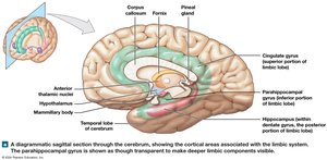
The limbic system is a functional grouping of brain structures that establishes emotional states, links conscious and autonomic functions, and facilitates memory storage and retrieval.
Components: Limbic lobe, hippocampus (learning and memory), amygdala (emotional responses and autonomic regulation).
The Cerebrum
Anatomy and Structure
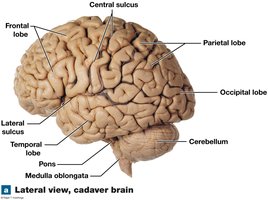
The cerebrum is the largest part of the brain, responsible for conscious thought, intellectual functions, and processing somatic sensory and motor information. It consists of two hemispheres, gray matter (cortex and basal nuclei), and white matter (deep to cortex).
Cerebral Cortex: Outer layer; contains gyri (elevations) and sulci (grooves).
Longitudinal Fissure: Separates hemispheres.
Functional Areas of the Cerebral Cortex
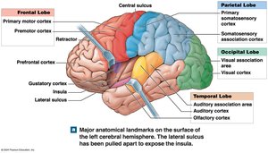
Primary Motor Cortex: Controls voluntary skeletal muscles; located in precentral gyrus (frontal lobe).
Primary Somatosensory Cortex: Receives touch, pressure, pain, vibration, and temperature; located in postcentral gyrus (parietal lobe).
Special Sensory Cortices: Visual (occipital lobe), auditory (temporal lobe), olfactory (temporal lobe), gustatory (insula).
Association Areas: Interpret sensory data and coordinate motor responses (somatosensory, visual, auditory association areas).
Motor Association Area (Premotor Cortex): Coordinates learned movements.
Integrative Centers
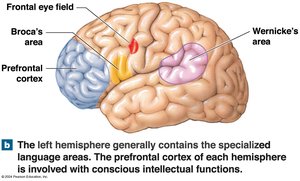
Prefrontal Cortex: Abstract intellectual functions, prediction, and analysis.
Wernicke's Area: Language comprehension; coordinates access to visual and auditory memories.
Broca's Area: Speech production; regulates breathing and vocalization.
Disorders: Aphasia (inability to speak/read), dyslexia (difficulty with written words).
Hemispheric Lateralization
Left Hemisphere: Language, math, decision making.
Right Hemisphere: Sensory analysis, recognition of faces and voice inflections.
Brain Waves and EEG
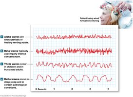
Brain activity can be visualized by PET or fMRI and assessed with EEG. Different types of brain waves are observed:
Alpha Waves: Healthy, awake adults at rest.
Beta Waves: Concentration and mental stress.
Theta Waves: Children and frustrated adults; may indicate disorder in adults.
Delta Waves: Sleep in infants; brain damage in awake adults.
Cranial Nerves
Classification and Origins
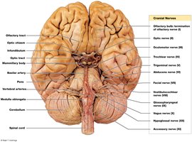
Cranial nerves are PNS nerves connected to the brain. They are classified as sensory, special sensory, motor, or mixed nerves.
Sensory: Touch, pressure, vibration, temperature, pain.
Special Sensory: Smell, sight, hearing, balance.
Motor: Somatic motor commands.
Mixed: Both sensory and motor fibers.
Summary Table: Major Brain Regions and Functions
Region | Main Function |
|---|---|
Cerebrum | Conscious thought, intellect, memory, sensation, complex movement |
Cerebellum | Coordination of movement, balance, equilibrium |
Diencephalon | Sensory relay, autonomic regulation, hormone production |
Brainstem | Relay between spinal cord and higher brain regions; autonomic functions |
Summary Table: Types of Brain Waves
Wave Type | Frequency | Associated State |
|---|---|---|
Alpha | 8–13 Hz | Awake, resting |
Beta | 13–30 Hz | Concentration, stress |
Theta | 4–7 Hz | Children, frustration |
Delta | 0.5–4 Hz | Deep sleep, brain damage |
Summary Table: Cranial Nerve Classifications
Type | Description |
|---|---|
Primarily Sensory | Somatic sensory information |
Special Sensory | Special senses (smell, sight, hearing, balance) |
Motor | Somatic motor commands |
Mixed | Both sensory and motor fibers |
Additional info: Tables were inferred and expanded for clarity and completeness. All images included are directly relevant to the adjacent content and reinforce anatomical and physiological concepts.
