 Back
BackChapter 14: The Brain and Cranial Nerves – Structured Study Notes
Study Guide - Smart Notes
 Tailored notes based on your materials, expanded with key definitions, examples, and context.
The Brain and Cranial Nerves
Introduction to the Brain and Cranial Nerves
The adult human brain is the central organ of the nervous system, containing nearly 97% of the body's nervous tissue. It weighs approximately 1.4 kg and has a volume of about 1200 mL, with normal variation between 750 and 2100 mL. Although male brains are about 10% larger than female brains, there is no correlation between brain size and intelligence.
Major Regions of the Brain
Overview of Brain Regions
Cerebrum: Largest part, responsible for higher mental functions such as thought, intellect, and memory.
Cerebellum: Coordinates repetitive body movements and maintains balance.
Diencephalon: Contains the thalamus and hypothalamus, involved in sensory processing, emotions, and hormone production.
Brainstem: Relays information between the spinal cord and higher brain regions; includes the midbrain, pons, and medulla oblongata.
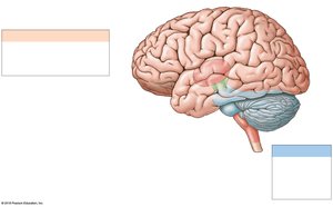
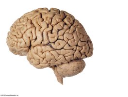
Cerebrum
Divided into left and right hemispheres.
Surface layer is the cerebral cortex (gray matter).
Gyri (ridges) increase surface area; separated by sulci (shallow grooves) and fissures (deep grooves).
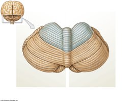
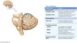
Cerebellum
Second-largest brain region.
Coordinates complex somatic motor patterns and adjusts output of other motor centers.
Covered by the cerebellar cortex (gray matter).
Diencephalon
Thalamus: Relays and processes sensory information.
Hypothalamus: Controls emotions, autonomic functions, and hormone production.
Pituitary gland: Major endocrine gland, connects to hypothalamus via the infundibulum.
Brainstem
Midbrain: Processes sight, sound, and associated reflexes; maintains consciousness.
Pons: Connects cerebellum to brainstem; contains nuclei for somatic and visceral motor control.
Medulla oblongata: Connects brain to spinal cord; regulates autonomic functions such as heart rate and digestion.
Development and Ventricles of the Brain
Embryonic Development
The brain develops from the neural tube, which forms three primary brain vesicles:
Prosencephalon (forebrain)
Mesencephalon (midbrain)
Rhombencephalon (hindbrain)
These differentiate into five secondary vesicles, which give rise to the major brain regions and ventricles.
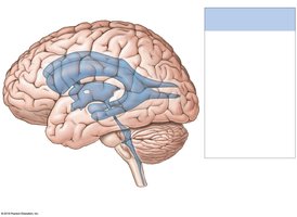
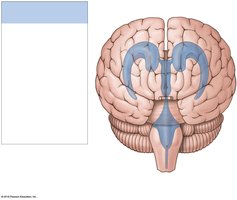
Ventricles of the Brain
Ventricles are fluid-filled chambers lined with ependymal cells.
Each cerebral hemisphere contains a lateral ventricle, separated by the septum pellucidum.
The third ventricle is in the diencephalon; the fourth ventricle extends into the medulla oblongata and connects with the central canal of the spinal cord.
Brain Protection and Support
Physical and Biochemical Protection
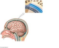
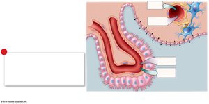
Protected by the cranium (skull bones), cranial meninges, and cerebrospinal fluid (CSF).
Biochemical isolation is provided by the blood-brain barrier (BBB).
Cranial Meninges
Three layers: dura mater (outer), arachnoid mater (middle), pia mater (inner).
Dural folds (falx cerebri, tentorium cerebelli, falx cerebelli) stabilize and support the brain.
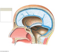
Cerebrospinal Fluid (CSF)
Produced by the choroid plexus (specialized ependymal cells and capillaries).
Functions: supports the brain, cushions neural structures, transports nutrients and wastes.
CSF circulates through ventricles, central canal, and subarachnoid space, and is absorbed into venous circulation via arachnoid granulations.
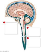
Blood-Brain Barrier (BBB) and Blood-CSF Barrier
BBB isolates CNS from general circulation; only lipid-soluble substances can diffuse freely.
Astrocytes regulate BBB permeability.
Blood-CSF barrier is formed by ependymal cells at the choroid plexus, allowing different composition of blood and CSF.
Some regions (hypothalamus, pituitary, pineal gland, choroid plexus) have breaks in the BBB for hormone release.
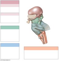
Brainstem: Medulla Oblongata, Pons, and Midbrain
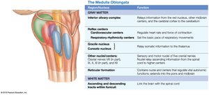
Medulla Oblongata
Most inferior part of the brainstem; connects brain to spinal cord.
Contains autonomic centers for cardiovascular and respiratory regulation.
Relay stations (gracile and cuneate nuclei, solitary nuclei, inferior olivary complex) process sensory and motor information.
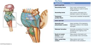
Pons
Contains sensory and motor nuclei for cranial nerves, nuclei involved in respiration (apneustic and pneumotaxic centers), and tracts that relay information to/from the cerebellum.
Transverse pontine fibers connect the pons to the cerebellum.
Midbrain
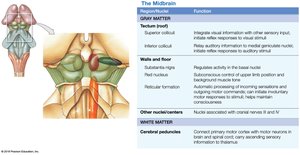
Tectum: Contains corpora quadrigemina (superior colliculi for visual reflexes, inferior colliculi for auditory reflexes).
Tegmentum: Contains red nucleus and substantia nigra (involved in motor control).
Cerebral peduncles: Contain descending motor fibers.
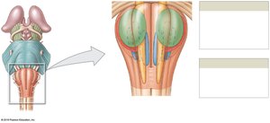
The Cerebellum
Structure and Function
Consists of cerebellar cortex (gray matter), folia (folds), anterior and posterior lobes, vermis (midline), and flocculonodular lobe.
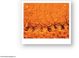
Purkinje cells in the cortex receive extensive synaptic input.
Arbor vitae is the internal white matter; cerebellar nuclei relay information to Purkinje cells.
Functions: adjusts postural muscles, fine-tunes movements, and maintains balance.
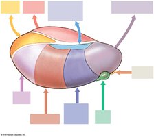
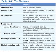
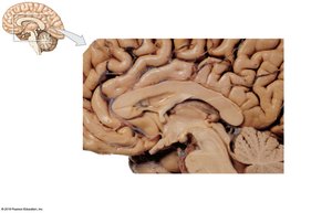
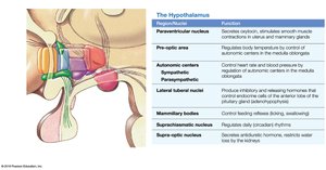
The Diencephalon
Thalamus
Major relay and processing center for sensory information.
Composed of several nuclei, each with specific sensory or motor relay functions.
Geniculate bodies relay visual and auditory information.
Hypothalamus
Regulates autonomic functions, emotions, hormone production, and circadian rhythms.
Connects to the pituitary gland via the infundibulum.
Contains centers for hunger, thirst, body temperature, and emotional responses.
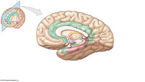
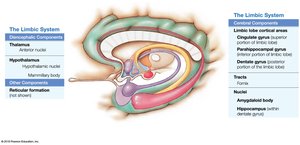
The Limbic System
Structure and Function
Establishes emotional states, links conscious and unconscious functions, and facilitates memory storage and retrieval.
Includes the limbic lobe, hippocampus, amygdaloid body, fornix, and anterior nuclei of the thalamus.
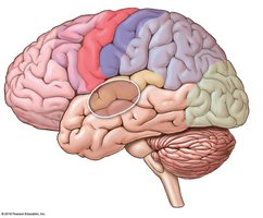
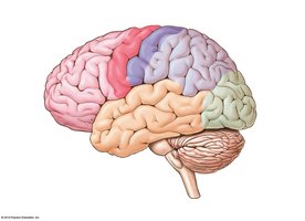
The Cerebrum
Structure
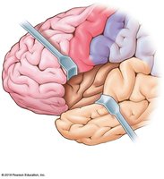
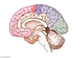
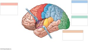
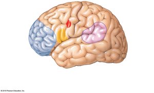
Divided into lobes: frontal, parietal, temporal, occipital, and insula.
Gyri and sulci increase surface area for cortical neurons.
Central sulcus separates frontal and parietal lobes; lateral sulcus separates frontal from temporal lobe; parieto-occipital sulcus separates parietal from occipital lobe.
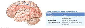
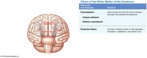
White Matter of the Cerebrum
Association fibers: Connect areas within the same hemisphere.
Commissural fibers: Connect the two hemispheres (e.g., corpus callosum).
Projection fibers: Link cortex with lower brain regions and spinal cord.
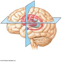
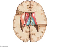
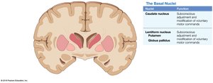
Basal Nuclei
Masses of gray matter deep within the cerebrum.
Include caudate nucleus, lentiform nucleus (putamen and globus pallidus), and claustrum.
Direct subconscious activities and coordinate learned movement patterns.
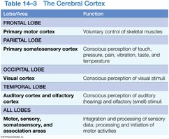
Functional Areas of the Cerebral Cortex
Primary motor cortex: Controls voluntary movements (precentral gyrus).
Primary somatosensory cortex: Receives sensory information (postcentral gyrus).
Special sensory cortices: visual, auditory, olfactory, and gustatory.
Association areas interpret sensory input and coordinate responses.
Hemispheric Lateralization
Left hemisphere: language, math, logic.
Right hemisphere: spatial analysis, recognition of faces, sensory analysis.
Brain Waves and EEG
EEG records electrical activity (brain waves): alpha, beta, theta, delta.
Alpha: resting adults; Beta: concentration; Theta: children/frustration; Delta: deep sleep/brain damage.
Cranial Nerves
Overview
12 pairs, classified as sensory, motor, or mixed.
Some distribute autonomic fibers to peripheral ganglia.
Selected Cranial Nerves
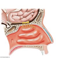
Olfactory (I): Special sensory (smell); origin: olfactory epithelium; destination: olfactory bulbs.
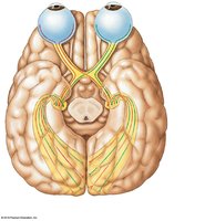
Optic (II): Special sensory (vision); origin: retina; destination: thalamus via optic chiasm.
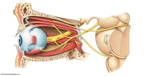
Oculomotor (III), Trochlear (IV), Abducens (VI): Motor nerves controlling eye movements.
Trigeminal (V): Mixed; sensory from face, motor to muscles of mastication.
Facial (VII): Mixed; sensory from taste receptors, motor to facial muscles and glands.
Vestibulocochlear (VIII): Special sensory; balance and hearing.
Glossopharyngeal (IX), Vagus (X), Accessory (XI), Hypoglossal (XII): Mixed or motor; involved in swallowing, taste, visceral organ control, and tongue movements.
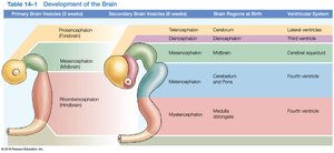
Summary Table: Development of the Brain
Primary Brain Vesicle | Secondary Vesicle | Brain Region at Birth | Ventricular System |
|---|---|---|---|
Prosencephalon | Telencephalon | Cerebrum | Lateral ventricles |
Prosencephalon | Diencephalon | Diencephalon | Third ventricle |
Mesencephalon | Mesencephalon | Midbrain | Cerebral aqueduct |
Rhombencephalon | Metencephalon | Cerebellum and Pons | Fourth ventricle |
Rhombencephalon | Myelencephalon | Medulla oblongata | Fourth ventricle |
Additional info: This table summarizes the embryological origins of the major brain regions and their associated ventricles.
Key Terms and Concepts
Gyri: Elevated ridges on the cerebral cortex.
Sulci: Shallow grooves separating gyri.
Fissures: Deep grooves in the brain.
Choroid plexus: Network producing CSF.
Basal nuclei: Subcortical gray matter involved in motor control.
Blood-brain barrier: Selective barrier protecting the CNS.
