 Back
BackChapter 14: The Somatic Nervous System – Sensory and Motor Pathways
Study Guide - Smart Notes
 Tailored notes based on your materials, expanded with key definitions, examples, and context.
Sensory Perception
Introduction to Sensory Perception
Sensory perception is the process by which sensory receptors convert environmental or internal stimuli into electrochemical signals, known as action potentials. These signals are then relayed to the central nervous system (CNS), where they may become conscious sensations or remain subconscious responses.
Sensory receptors detect changes in the environment or within the body.
Action potentials are the electrochemical signals generated by sensory neurons in response to stimuli.
Perception is the conscious awareness of a sensation, such as smelling cookies.
Sensation refers to the activation of sensory receptor cells, which may or may not reach conscious awareness.
Many internal stimuli are processed subconsciously, such as those from internal organs.
Sensory Receptors
Types of Sensory Receptors
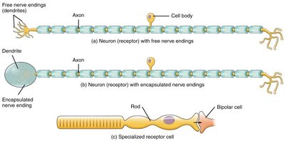
Sensory receptors are specialized cells or structures that detect specific types of stimuli. They can be classified based on their structure and the type of stimulus they detect.
Free nerve endings: Dendrites of sensory neurons embedded directly in tissues. Example: Nociceptors (pain receptors) and thermoreceptors (temperature receptors) in the skin.
Encapsulated nerve endings: Sensory neurons with dendrites enclosed in connective tissue. Example: Mechanoreceptors for pressure and touch in the skin.
Specialized receptor cells: Non-neuronal cells that synapse with sensory neurons. Example: Photoreceptors in the retina for light detection.
Chemoreceptors: Detect chemical stimuli, such as odorants (smell) and tastants (taste).
Sensory Pathways
Organization of Sensory Pathways
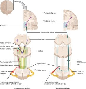
Sensory information from the body is transmitted to the CNS via spinal nerves, which split into dorsal and ventral roots near the spinal cord. The dorsal root contains only sensory neuron axons, with cell bodies located in the dorsal root ganglion. Sensory information ascends to the brain through specific tracts, typically involving three consecutive neurons.
Dorsal root: Contains sensory neuron axons; cell bodies are in the dorsal root ganglion.
Ascending tracts: Pathways that carry sensory information to the brain.
Most sensory pathways are contralateral, meaning the right side of the body is connected to the left side of the brain and vice versa.
Decussation: The crossing over of nerve fibers to the opposite side of the CNS; the location of decussation varies by pathway.
Cortical Processing
Somatosensory Cortex and Sensory Homunculus
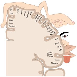
The primary somatosensory cortex, located in the postcentral gyrus of the parietal lobe, is responsible for processing sensory information such as touch, proprioception, pain (nociception), and temperature. The sensory homunculus is a visual representation of the cortical area dedicated to processing sensory input from different body regions. More sensitive areas are depicted as larger in the homunculus.
Primary somatosensory cortex: Receives and processes sensory input.
Sensory homunculus: Illustrates the proportional representation of body regions in the cortex.
Descending Pathways
Motor Pathways and the Corticospinal Tract
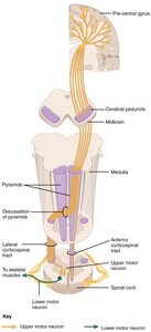
Motor commands from the brain are transmitted to skeletal muscles via descending pathways. The ventral roots of spinal nerves contain axons of motor neurons, whose cell bodies are in the ventral horns of the spinal cord. The corticospinal tract is the primary descending pathway for voluntary motor control, involving two consecutive neurons: the upper motor neuron (originating in the brain) and the lower motor neuron (projecting to skeletal muscle).
Corticospinal tract: Main pathway for voluntary motor control.
Upper motor neuron: Originates in the cerebral cortex and synapses in the spinal cord.
Lower motor neuron: Projects from the spinal cord to skeletal muscle, ending at the neuromuscular junction.
Most motor fibers are contralateral, crossing over to the opposite side of the body.

Pyramids and Decussation
The pyramids are structures in the medulla oblongata composed of white matter, containing motor fibers of the corticospinal tract. Most fibers decussate (cross over) at the pyramidal decussation, forming the lateral corticospinal tract (controlling appendicular muscles). Some fibers decussate within the spinal cord, forming the anterior corticospinal tract (controlling trunk muscles). A minority of anterior corticospinal tract fibers remain ipsilateral.
Pyramidal decussation: Site where most corticospinal fibers cross to the opposite side.
Lateral corticospinal tract: Controls limb muscles; fibers decussate in the medulla.
Anterior corticospinal tract: Controls trunk muscles; some fibers decussate in the spinal cord, others remain ipsilateral.
Summary Table: Types of Sensory Receptors
Receptor Type | Structure | Stimulus Detected | Example |
|---|---|---|---|
Free nerve ending | Unencapsulated dendrite | Pain, temperature | Nociceptor, thermoreceptor |
Encapsulated nerve ending | Dendrite enclosed in connective tissue | Pressure, touch | Mechanoreceptor |
Specialized receptor cell | Non-neuronal cell synapsing with neuron | Light, chemicals | Photoreceptor, chemoreceptor |
Additional info: The somatic nervous system is responsible for voluntary control of body movements via skeletal muscles and for processing sensory information from the external environment. Understanding the organization of sensory and motor pathways is essential for comprehending how the nervous system integrates and responds to stimuli.
