 Back
BackChapter 15: Sensory Pathways and the Somatic Nervous System – Study Notes
Study Guide - Smart Notes
 Tailored notes based on your materials, expanded with key definitions, examples, and context.
Chapter 15: Sensory Pathways and the Somatic Nervous System
Introduction
This chapter explores the organization and function of sensory and motor pathways in the nervous system, focusing on how the body detects, processes, and responds to sensory information. It also covers the structure and function of general and special sensory receptors, the organization of sensory and motor tracts, and the integration of sensory input and motor output.
15-1 Sensory and Motor Pathways
Overview of Sensory and Motor Pathways
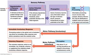
Sensory pathways are series of neurons that relay sensory information from sensory receptors to the central nervous system (CNS).
Sensory receptors are specialized cells or neuron processes that monitor specific conditions in the body or external environment.
When stimulated, receptors generate action potentials that travel along sensory pathways to the CNS.
The afferent division of the nervous system includes somatic and visceral sensory pathways.
Somatic sensory information is processed in the cerebral cortex; visceral sensory information is processed mainly in the brainstem and diencephalon.
The efferent division includes somatic motor pathways that control skeletal muscles (effectors).
Motor commands originate in the brain and travel along somatic motor pathways, potentially modified by higher-order brain functions.
15-2 Sensory Receptors
Sensation and Perception
Sensation: Sensory information arriving in the CNS.
Perception: Conscious awareness of a sensation.
Types of Senses
General senses: Temperature, pain, touch, pressure, vibration, proprioception (body position).
Special senses: Olfaction (smell), gustation (taste), vision (sight), equilibrium (balance), hearing.
Special sensory receptors are located in sense organs such as the eye or ear.
Transduction and Receptor Specificity
Transduction: Conversion of a stimulus into an action potential by a sensory receptor.
Receptor specificity: Each receptor has a characteristic sensitivity to a particular stimulus.
Receptive field: The area monitored by a single receptor cell. Larger receptive fields make it harder to localize stimuli.

Receptor Potentials and Adaptation
Receptor potential: Change in membrane potential of the receptor due to a stimulus; can be depolarizing (generator potential) or hyperpolarizing.
The size of the receptor potential depends on stimulus strength.
Adaptation: Reduction in receptor sensitivity during constant stimulation.
Peripheral adaptation occurs in the PNS; central adaptation occurs in the CNS.
Fast-adapting receptors (phasic) respond strongly at first, then decrease activity (e.g., temperature receptors).
Slow-adapting receptors (tonic) show little adaptation (e.g., pain receptors).
Tonic and Phasic Receptors
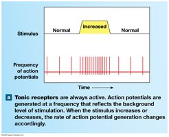
Tonic receptors: Always active; frequency of action potentials reflects the level of stimulation.
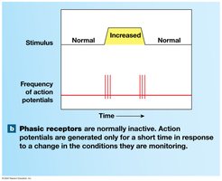
Phasic receptors: Normally inactive; generate action potentials only in response to changes in the monitored condition.
15-3 General Sensory Receptors
Classification by Location
Exteroceptors: Monitor the external environment.
Proprioceptors: Monitor position of skeletal muscles and joints.
Interoceptors: Monitor visceral organs and functions.
Classification by Stimulus Type
Nociceptors: Detect pain.
Thermoreceptors: Detect temperature.
Mechanoreceptors: Detect physical distortion (touch, pressure, vibration).
Chemoreceptors: Detect chemical concentrations.
Nociceptors (Pain Receptors)
Free nerve endings with large receptive fields; common in skin, joint capsules, periosteum, and blood vessel walls.
Sensitive to temperature extremes, mechanical damage, or chemicals from injured cells.
Tonic receptors with little peripheral adaptation; central adaptation can reduce pain perception.
Endorphins and enkephalins inhibit pain pathways in the CNS.
Types of Pain
Fast pain: Sharp, prickling pain; carried by myelinated Type A fibers; triggers reflexes and conscious awareness.
Slow pain: Burning, aching pain; carried by unmyelinated Type C fibers; causes generalized awareness.
Thermoreceptors
Free nerve endings in dermis, skeletal muscles, liver, and hypothalamus.
Phasic receptors; respond to temperature changes, then adapt.
Pathways are similar to those for pain sensations.
Mechanoreceptors
Sensitive to physical distortion of their plasma membranes.
Contain mechanically-gated ion channels that open/close with stretching, compression, or twisting.
Three main classes: Tactile receptors (touch, pressure, vibration), Baroreceptors (pressure), Proprioceptors (position).
Tactile Receptors in the Skin
Free nerve endings: Tonic receptors for touch, pressure, pain, and temperature; small receptive fields.

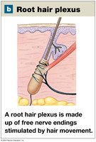
Root hair plexus: Nerve endings that detect hair movement; adapt rapidly.
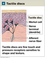
Tactile discs: Sensitive tonic receptors for fine touch and pressure; small receptive fields.
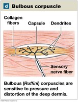
Bulbous corpuscles (Ruffini): Tonic receptors for pressure and skin distortion; located in deep dermis; little adaptation.
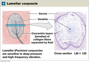
Lamellar corpuscles (Pacinian): Fast-adapting receptors for deep pressure and high-frequency vibration; single dendrite in concentric collagen layers.
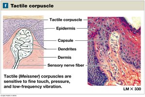
Tactile corpuscles (Meissner): Large, fast-adapting receptors for fine touch, pressure, and low-frequency vibration; abundant in sensitive areas (eyelids, lips, fingertips, nipples, external genitalia).
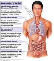
Baroreceptors
Detect pressure changes in blood vessels and organs of the digestive, respiratory, and urinary tracts.
Free nerve endings that branch within elastic tissues; adapt rapidly to pressure changes.
Proprioceptors
Monitor position of joints and skeletal muscles; only somatic sensation (not found in visceral organs).
Types: Muscle spindles (muscle length, stretch reflexes), Golgi tendon organs (tension during contraction), Joint capsule receptors (pressure, tension, movement).
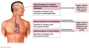
Chemoreceptors
Respond to water- and lipid-soluble substances dissolved in body fluids.
Exhibit fast peripheral adaptation.
Monitor pH, CO2, and O2 levels in arterial blood (carotid and aortic bodies).
15-4 Sensory Pathways
Organization of Sensory Pathways
First-order neuron: Delivers sensations from receptors to the CNS; cell body in spinal or cranial nerve ganglion.
Second-order neuron: Interneuron in spinal cord or brainstem; axon crosses to opposite side (decussation).
Third-order neuron: Located in the thalamus; relays information to the primary somatosensory cortex.
Somatic Sensory Pathways
Carry sensory information from skin, muscles, head, neck, and limbs to the CNS.
Three main pathways: Spinothalamic, Posterior column, Spinocerebellar.
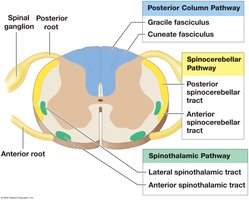
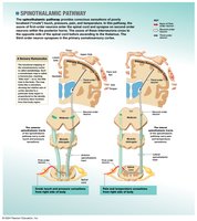
Spinothalamic Pathway
Carries sensations of crude touch, pressure, pain, and temperature.
First-order neurons synapse in posterior horns; second-order neurons cross and ascend to thalamus; third-order neurons project to somatosensory cortex.
Perception depends on which neurons are stimulated and where the thalamus sends information.
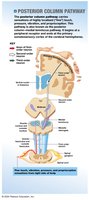
Posterior Column Pathway
Carries sensations of fine touch, vibration, pressure, and proprioception.
First-order neurons ascend to medulla oblongata; second-order neurons decussate and ascend to thalamus; third-order neurons project to somatosensory cortex.
Sensory Homunculus
Functional map of the primary somatosensory cortex.
Area devoted to a body region is proportional to sensory neuron density, not region size (e.g., large for lips, small for back).
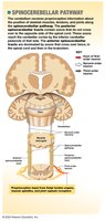
Spinocerebellar Pathway
Carries information about positions of muscles, tendons, and joints to the cerebellum.
First-order neurons synapse in posterior horn; second-order neurons ascend to cerebellum, often decussating twice.
Information does not reach conscious awareness.
Visceral Sensory Pathways
Interoceptors monitor visceral tissues and organs in thoracic and abdominopelvic cavities.
First-order neurons in cranial and spinal nerves; second-order interneurons ascend to solitary nuclei of medulla oblongata.
Solitary nuclei connect with cardiovascular, respiratory centers, and reticular formation.
15-5 Somatic Motor Pathways
Organization of Somatic Motor Pathways
Somatic nervous system (SNS) controls skeletal muscles.
Somatic motor pathways always involve at least two motor neurons:
Upper motor neuron: Cell body in CNS processing center (primary or premotor cortex); axon synapses on lower motor neuron.
Lower motor neuron: Cell body in brainstem or spinal cord; axon extends to skeletal muscle.
Major Motor Pathways
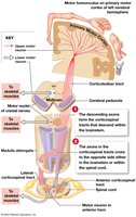
Corticospinal pathway (pyramidal system): Provides voluntary control over skeletal muscles; upper motor neurons are pyramidal cells of the primary motor cortex.
Medial pathway: Controls muscle tone and gross movements of trunk and proximal limbs.
Lateral pathway: Controls muscle tone and precise movements of distal limbs.
Basal nuclei and cerebellum monitor and adjust these pathways.
Motor Homunculus
Functional map of the primary motor cortex; size of area corresponds to degree of fine motor control (large for hands, face, tongue).
Proportions are similar to the sensory homunculus.
Role of Basal Nuclei and Cerebellum
Basal nuclei: Provide background patterns of movement for voluntary activities; can stimulate or inhibit upper motor neurons.
Influence premotor cortex and reticular formation to alter motor output.
Cerebellum: Monitors proprioceptive, visual, and vestibular information to adjust movement; inhibits unnecessary motor commands; patterns are learned by repetition.
Summary Table: Types of Sensory Receptors
Receptor Type | Stimulus Detected | Location | Adaptation |
|---|---|---|---|
Nociceptors | Pain (mechanical, thermal, chemical) | Skin, joints, periosteum, blood vessels | Slow (tonic) |
Thermoreceptors | Temperature | Dermis, muscles, liver, hypothalamus | Fast (phasic) |
Mechanoreceptors | Touch, pressure, vibration, stretch | Skin, muscles, organs | Varies |
Chemoreceptors | Chemical concentration (pH, CO2, O2) | Carotid/aortic bodies, CNS | Fast |
Key Equations
Action Potential Generation:
Receptor Potential:
Learning Outcomes
Specify the components of the afferent and efferent divisions of the nervous system and explain the somatic nervous system.
Explain why receptors respond to specific stimuli and how receptor organization affects sensitivity.
Identify receptors for general senses and describe their function.
Identify major sensory pathways and explain how sensations from different body areas are distinguished.
Describe the components, processes, and functions of somatic motor pathways and levels of motor control.
