Back
BackChapter 15: The Special Senses – Anatomy & Physiology Study Notes
Study Guide - Smart Notes
 Tailored notes based on your materials, expanded with key definitions, examples, and context.
Module 15.1 Comparison of General and Special Senses
Overview of Sensory Systems
The human body detects environmental changes through general and special senses, each with distinct anatomical and physiological features.
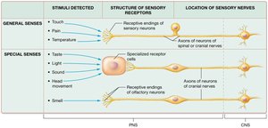
General senses detect touch, pain, and temperature using sensory neuron endings distributed throughout the body.
Special senses (smell, taste, vision, hearing, vestibular sensation) use specialized organs located in the head and are innervated by cranial nerves.
Special senses detect specific stimuli: light, sound waves, head movements, and chemicals.
Sensory Transduction
Transduction is the process by which physical or chemical stimuli are converted into action potentials interpretable by the brain.
General sensory neurons have specialized receptive endings that alter their membrane potential in response to stimuli.
Special senses use receptor cells (except olfaction, which uses neurons) to transduce stimuli into action potentials.
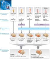
Sensory signals are relayed to the thalamus and primary sensory cortices for awareness, then to association areas for integration.
Module 15.2 Anatomy and Physiology of Smell (Olfaction)
Structures of Olfaction
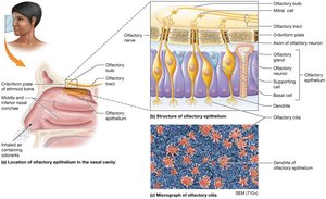
The olfactory system detects airborne chemicals (odorants) and transduces them into signals perceived as odors.
Olfactory epithelium: Located in the superior nasal cavity; contains three cell types:
Olfactory neurons: Modified bipolar neurons (chemoreceptors) that detect odorants.
Basal cells: Stem cells that replace olfactory neurons every 30–60 days.
Supporting cells: Columnar cells that surround and support olfactory neurons.
Olfactory nerve (CN I): Formed by axons of olfactory neurons.
Olfactory bulb: Sits above the cribriform plate; receives input from olfactory neurons.
Olfactory tract: Axons from the bulb travel to the CNS for interpretation.
Module 15.3 Anatomy and Physiology of Taste (Gustation)
The Gustatory Sense
Taste is detected by chemoreceptors in taste buds, with input from olfactory, thermal, and pain receptors.
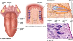
Taste buds: Clusters of receptor and supporting cells on the tongue and oral cavity surfaces.
Each taste bud is associated with a sensory neuron that transmits information to the CNS.
Structures of Gustation
Papillae: Projections on the tongue classified by shape:
Vallate (circumvallate): Dome-shaped, contain hundreds of taste buds.
Fungiform: Mushroom-shaped, contain few taste buds.
Foliate: Ridges on tongue sides, taste buds present in childhood.
Filiform: Long, thin, no taste buds; detect texture and temperature.
Taste buds contain:
Gustatory cells: Epithelial cells with microvilli (taste hairs) that detect chemicals.
Basal cells: Stem cells for gustatory cell replacement (lifespan 10–14 days).
Supporting cells: Physically support gustatory cells.
Physiology of Gustation
Taste involves transduction of chemicals into electrical signals. Five primary taste sensations:
Sweet: Simple sugars (e.g., glucose, fructose).
Sour: Hydrogen ions (e.g., citric acid).
Salty: Metal ions (e.g., sodium, potassium).
Bitter: Nitrogen-containing compounds (often toxic).
Umami: Amino acids (e.g., glutamate; savory taste).
Module 15.4 Anatomy of the Eye
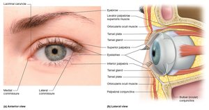
Accessory Structures of the Eye
The eye is protected and maintained by several accessory structures within the orbit.
Eyelids (palpebrae): Protect the eye, distribute tears, and prevent foreign object entry.
Tarsal plates/glands: Provide structure and secrete oil to prevent eyelid adhesion.
Medial/lateral commissures: Where eyelids meet; contain the lacrimal caruncle (secretes lubricating substance).
Muscles: Orbicularis oculi (closes eyelid), levator palpebrae superioris (opens eyelid).
Eyebrows/eyelashes: Protect from debris and light, trigger blink reflex.
Conjunctiva: Thin membrane lining eyelids and covering the sclera.
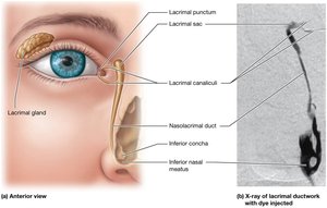
Lacrimal Apparatus
Lacrimal gland: Produces tears for lubrication and debris removal.
Tears drain through lacrimal puncta, canaliculi, sac, and nasolacrimal duct into the nasal cavity.
Extrinsic Eye Muscles
Six muscles control eye movement: superior, inferior, lateral, and medial rectus; superior and inferior oblique.
Innervation: CN IV (superior oblique), CN VI (lateral rectus), CN III (others).
Mnemonic: "LR6SO4 all the rest are III".
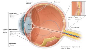
Structure of the Eyeball
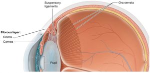
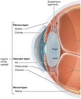
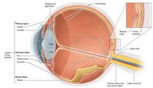
The eyeball is a hollow sphere with three main tissue layers:
Fibrous layer: Outermost; includes sclera (white, protective) and cornea (transparent, refracts light).
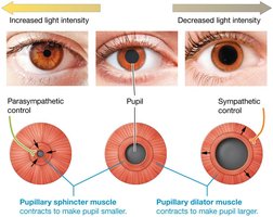
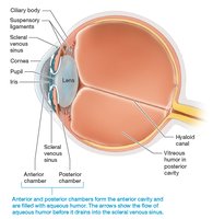
Vascular layer: Middle; includes choroid (vascular, pigmented), ciliary body (muscle, controls lens), iris (colored part, controls pupil size), and pupil (light entry).
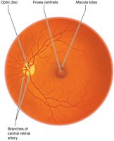
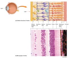
Neural layer (retina): Innermost; contains photoreceptors (rods and cones), macula lutea, fovea centralis (sharp vision), and optic disc (blind spot).
Cavities and Chambers of the Eye
Posterior cavity: Behind lens; filled with vitreous humor (maintains shape, presses retina against choroid).
Anterior cavity: In front of lens; contains aqueous humor (nourishes cornea/lens, drains via scleral venous sinus).
Module 15.5 Anatomy and Physiology of Vision
Principles of Light
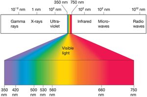
Vision is based on the detection of light, a form of electromagnetic radiation.
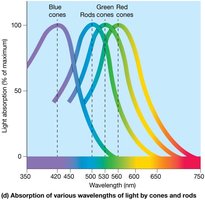
Visible light: 350–750 nm wavelength; shorter wavelengths are blue/violet, longer are red.
Photon: Basic unit of light, stimulates retinal photoreceptors.
Refraction of Light
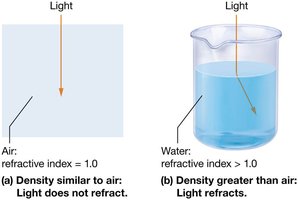
Light bends (refracts) when passing through substances of different densities (refractive index).
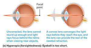
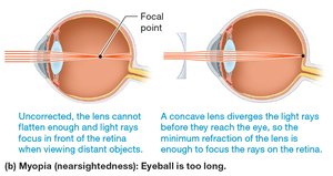
Convex lenses converge light; concave lenses diverge it.
Focusing Light on the Retina
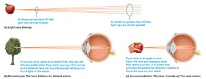
Cornea provides most refraction; lens fine-tunes focus.
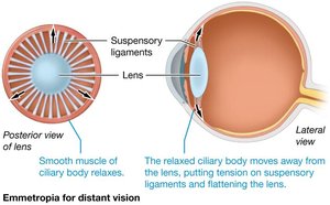
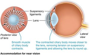
Accommodation: Lens thickens for near vision, flattens for distance.
Pupillary constriction and convergence aid near focus.
Errors of Refraction
Hyperopia (farsightedness): Eyeball too short/cornea too flat; corrected with convex lenses.
Myopia (nearsightedness): Eyeball too long/cornea too curved; corrected with concave lenses.
Astigmatism: Irregular curvature; corrected with special lenses or LASIK surgery.
Photoreceptors and the Retina
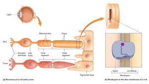
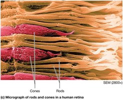
Rods: Black/white vision, low light, peripheral vision; contain rhodopsin (opsin + retinal).
Cones: Color vision, high acuity, bright light; contain iodopsin (photopsin + retinal).
Photoreceptors synapse with bipolar cells, which communicate with ganglion cells (form optic nerve).
The Visual Pathway
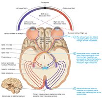
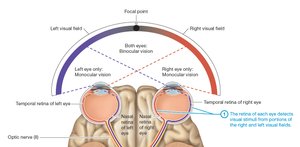
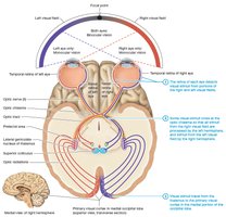
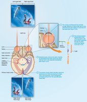
Retina detects visual stimuli; some axons cross at the optic chiasma.
Optic tracts carry information to the lateral geniculate nucleus (thalamus), then to the primary visual cortex.
Visual processing involves dorsal (motion) and ventral (form/color) pathways.
Module 15.6 Anatomy of the Ear
Regions of the Ear
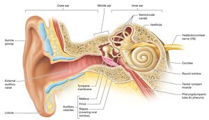
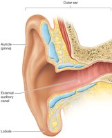
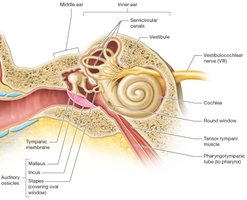
The ear is divided into external, middle, and inner regions, each with specialized structures for hearing and equilibrium.
External ear: Auricle (funnels sound), external auditory canal (conducts sound), tympanic membrane (vibrates with sound).
Middle ear: Air-filled chamber with auditory ossicles (malleus, incus, stapes) that amplify sound; pharyngotympanic tube equalizes pressure.
Inner ear: Bony and membranous labyrinths; contains cochlea (hearing), vestibule, and semicircular canals (equilibrium).
Inner Ear Structure
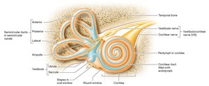
Bony labyrinth: Contains perilymph (extracellular-like fluid).
Membranous labyrinth: Contains endolymph (intracellular-like fluid).
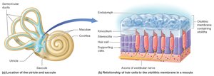
Vestibule: Contains utricle and saccule (detect head position, linear movement).
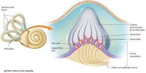
Semicircular canals: Detect rotational movement; ampullae contain hair cells.
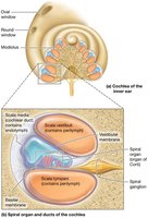
Cochlea: Contains cochlear duct (endolymph), scala vestibuli and tympani (perilymph), spiral organ (hair cells for hearing).
Module 15.7 Physiology of Hearing
Sound Transduction in the Inner Ear
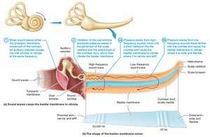
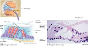
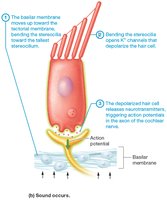
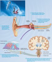
Sound waves vibrate the basilar membrane, moving hair cells of the spiral organ.
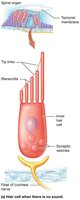
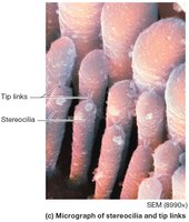
Stereocilia bend, opening potassium channels and depolarizing hair cells (unique to inner ear physiology).
Depolarization triggers neurotransmitter release and action potentials in the cochlear nerve (CN VIII).
Pitch is determined by which part of the basilar membrane vibrates; loudness by the amplitude of vibration.
Module 15.8 Anatomy and Physiology of Vestibular Sensation
Vestibular System and Equilibrium
Equilibrium depends on input from the visual system, proprioceptors, and vestibular system (utricle, saccule, semicircular canals).
Static equilibrium: Maintains balance when head is tilted but not moving.
Dynamic equilibrium: Maintains balance during movement (rotational, angular, or linear acceleration).
Utricle and Saccule
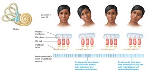
Contain maculae with hair cells embedded in the otolithic membrane (contains otoliths).
Bending of stereocilia alters glutamate release, changing action potential frequency in vestibular nerve.
Detect head tilt and linear acceleration.
Semicircular Ducts
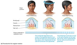
Detect angular and rotational movements.
Each ampulla contains a crista ampullaris (hair cells in cupula).
Movement of endolymph bends cupula, altering glutamate release and vestibular nerve activity.
Module 15.9 How the Special Senses Work Together
Sensory Integration
Special senses collect and transmit information simultaneously for integration in the brain.
Signals are relayed through the thalamus (except olfaction) to primary sensory cortices.
Frontal lobe and limbic system integrate sensory input, emotional response, and memory formation.