 Back
BackChapter 15: The Special Senses – Human Anatomy & Physiology Study Notes
Study Guide - Smart Notes
 Tailored notes based on your materials, expanded with key definitions, examples, and context.
Chapter 15: The Special Senses
15.1 Overview of the Special Senses
The special senses are those that convey specific stimuli from specialized sensory organs located in discrete regions of the head. These senses include smell (olfaction), taste (gustation), vision, hearing (audition), and vestibular sensation (balance). They are distinct from the general senses, which detect touch, pain, and temperature throughout the body.
Special Senses: Smell, taste, vision, hearing, and balance.
General Senses: Touch, pain, temperature.
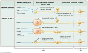
15.1 Comparison of the General and Special Senses
General senses detect a wide range of stimuli, while special senses detect very specific stimuli such as light, sound waves, head movements, and chemicals. The structure and location of sensory receptors differ between these two categories.
General Senses: Receptive ends of sensory neurons; stimuli transmitted via spinal and cranial nerves.
Special Senses: Specialized receptor cells (except olfaction, which uses modified neurons); all housed in the head and transmitted via cranial nerves.
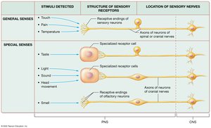
15.1 Sensory Transduction
Transduction is the process by which a physical or chemical stimulus is converted into an action potential interpretable by the brain. Special senses detect environmental stimuli and transduce them into action potentials, which are processed by sensory nuclei, the thalamus, and primary cortical areas for awareness and identification, then passed to association areas for interpretation and integration.
15.2 Olfaction (Smell)
Structures of Olfaction
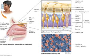
Olfaction allows detection of odorants (chemicals in the air). The olfactory epithelium is located in the superior region of each nasal cavity and contains three cell types:
Olfactory Neurons: Modified bipolar neurons with non-motile cilia; axons form the olfactory nerve.
Basal Cells: Stem cells that replace olfactory neurons (lifespan 30–60 days).
Supporting Cells: Columnar cells that support and surround olfactory neurons.
Physiology of Olfaction
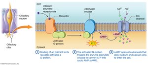
Odorants bind to odorant-binding proteins, which transport them to receptors on olfactory cilia. Binding activates a G-protein, which triggers adenylate cyclase to convert ATP into cyclic AMP (cAMP). cAMP opens ion channels, allowing sodium and calcium ions to enter, creating a local potential that depolarizes the membrane.
The Olfactory Pathway
Axons of olfactory neurons carry stimuli to the olfactory bulb, synapsing with mitral cells.
Stimuli travel from the olfactory bulb to the primary olfactory cortex in the temporal lobe (no thalamic relay).
Neurons from the olfactory cortex connect to the amygdala, hippocampus, hypothalamus, and limbic system, evoking emotional and visceral responses to odors.
15.3 Gustation (Taste)
Structures of Gustation: Taste Buds
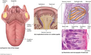
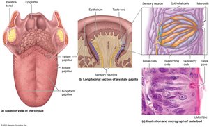
Gustation involves stimulation of specialized receptor cells in taste buds, which are found on the tongue and parts of the oral cavity. The tongue is covered with papillae:
Vallate (Circumvallate) Papillae: Large, dome-shaped, with hundreds of taste buds.
Fungiform Papillae: Mushroom-shaped, with a few taste buds.
Foliate Papillae: Ridges on the sides of the tongue, taste buds present only in childhood.
Filiform Papillae: Long, thin, lack taste buds but detect texture and temperature.
Taste buds contain:
Gustatory (Taste) Cells: Specialized epithelial cells with receptors on microvilli; synapse with sensory neurons of facial, glossopharyngeal, or vagus nerves.
Basal Cells: Stem cells for new gustatory cells.
Supporting Cells: Provide physical support.
Physiology of Gustation
Taste sensations depend on detection of chemicals and combinations of taste receptors:
Sweet: Simple sugars (e.g., glucose, fructose).
Sour: Hydrogen ions (e.g., citric acid).
Salty: Metal ions (e.g., sodium, potassium).
Bitter: Alkaloids (e.g., coffee, rancid foods); protective function.
Umami: Glutamate or other amino acids (savory).
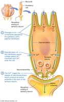
Activation of taste receptors requires liquid (usually saliva) to reach taste buds. Ion movement depolarizes the gustatory cell membrane, opening voltage-gated calcium channels. Calcium triggers neurotransmitter release, producing an action potential in the sensory neuron.
The Gustatory Pathway
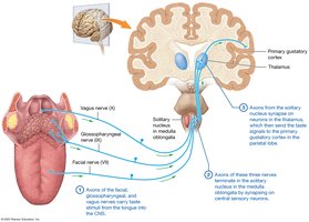
Facial, glossopharyngeal, and vagus nerves carry taste stimuli to the CNS.
Axons terminate in the solitary nucleus (medulla oblongata), then synapse in the thalamus.
Thalamic neurons project to the primary gustatory cortex (parietal lobe), insula, and inferior frontal lobe for integration with visual and olfactory stimuli.
15.4 The Eye and Vision
Accessory Structures of the Eye
Accessory structures support, protect, or move the eyeball:
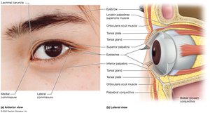
Eyelids (Palpebrae): Cover the anterior orbit, prevent foreign objects from entering, distribute tears.
Eyebrows: Prevent perspiration from entering eyes, reduce glare, aid facial expression.
Eyelashes: Trigger blink reflex.
Conjunctiva: Thin membrane lining eyelids and covering anterior eye; inflammation is conjunctivitis (“pink eye”).
Lacrimal Apparatus
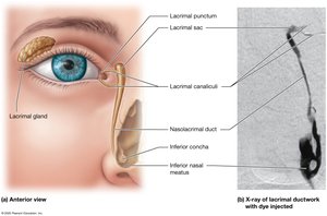
The lacrimal apparatus produces and drains tears, lubricating and cleaning the eye. Tears are produced by the lacrimal gland, drain through lacrimal puncta, canaliculi, sac, and nasolacrimal duct to the nasal cavity.
The Eyeball: Structure and Layers
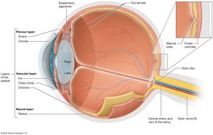
The eyeball is a hollow sphere with three tissue layers (tunics):
Fibrous Layer: Outermost; includes sclera (white, maintains shape) and cornea (transparent, allows light entry).
Vascular Layer: Middle; includes choroid (blood supply, pigment), ciliary body (smooth muscle, lens shape), and iris (pigmented, controls pupil size).
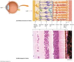
Neural Layer (Retina): Innermost; contains photoreceptors (rods and cones), bipolar cells, ganglion cells, and forms the optic nerve.
Lens and Chambers
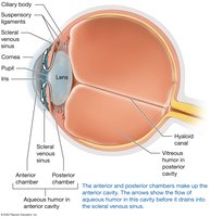
The lens focuses light on the retina. The posterior cavity contains vitreous humor (gelatinous, maintains shape), while the anterior cavity contains aqueous humor (fluid, nourishes cornea and lens, drains via scleral venous sinus).
Common Eye Disorders
Cataracts: Clouded lens, leading cause of blindness; treated by lens replacement.
Glaucoma: Increased intraocular pressure damages retina and optic nerve; treated with medication or surgery.


15.5 Principles of Light and Vision
Nature of Light
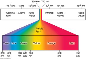
Light is electromagnetic radiation with wavelengths measured in nanometers (nm). The visible spectrum ranges from about 350–750 nm. The basic unit is the photon, which stimulates retinal photoreceptors.
Refraction and Lenses
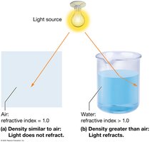
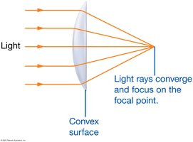
Light bends (refracts) when passing through materials of different densities. The refractive index quantifies this effect. Convex lenses converge light to a focal point; concave lenses diverge light.
Focusing Light on the Retina
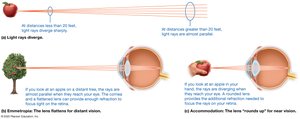
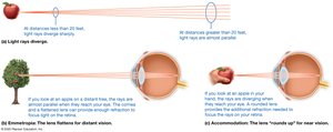
The cornea provides most of the eye’s refractive power; the lens fine-tunes focus. Distant objects require little lens adjustment (emmetropic state), while near objects require accommodation (lens rounds up).
Errors of Refraction
Presbyopia: Age-related loss of accommodation.
Astigmatism: Irregular curvature of lens or cornea.
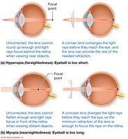
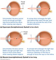
Hyperopia (Farsightedness): Eyeball too short; corrected with convex lenses.
Myopia (Nearsightedness): Eyeball too long; corrected with concave lenses.
15.5 Photoreceptors and the Retina
Retinal Structure
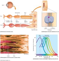
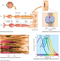
The retina contains rods (black and white, low light, peripheral vision) and cones (color, high acuity, bright light, central vision). Photoreceptors synapse with bipolar and ganglion cells, forming the optic nerve.
Photoreceptor Function
Rods: Contain rhodopsin (opsin + retinal); sensitive in dim light.
Cones: Contain iodopsin (photopsin + retinal); three types for blue, green, red wavelengths.
Phototransduction
In darkness, photoreceptors are depolarized and release neurotransmitters. Light hyperpolarizes photoreceptors, reducing neurotransmitter release and generating action potentials in ganglion cells.
15.6 Anatomy of the Ear and Hearing
Regions of the Ear
Outer Ear: Auricle (pinna), external auditory canal, tympanic membrane.
Middle Ear: Auditory ossicles (malleus, incus, stapes), pharyngotympanic tube.
Inner Ear: Bony and membranous labyrinths, cochlea (hearing), vestibule and semicircular canals (balance).
Transmission of Sound
Sound waves are funneled by the auricle, vibrate the tympanic membrane, and are amplified by ossicles to the oval window. Vibrations create pressure waves in cochlear fluids, moving the basilar membrane and stimulating hair cells in the spiral organ (Organ of Corti).
Auditory Pathway
Action potentials travel via the cochlear nerve to the cochlear nuclei (medulla-pons junction), then to the superior olivary nucleus, inferior colliculus, medial geniculate nucleus (thalamus), and finally to the primary auditory cortex (temporal lobe).
Hearing Loss
Conduction Hearing Loss: Problem in outer/middle ear (e.g., cerumen, infection, ossicle fusion).
Sensorineural Hearing Loss: Defect in cochlea or neural pathways (e.g., hair cell dysfunction, nerve damage).
15.8 Vestibular Sensation (Balance)
Vestibular System
The vestibular system (utricle, saccule, semicircular canals) detects head position and movement, providing equilibrium. The utricle and saccule detect static equilibrium and linear acceleration; semicircular ducts detect rotational equilibrium.
Vestibular Pathway
Action potentials propagate to vestibular nuclei (medulla-pons junction), then to the thalamus, parietal lobe, cranial nerve nuclei (eye movement), cerebellum, and spinal cord (muscle coordination).
Summary Table: General vs. Special Senses
Type | Stimuli Detected | Structure of Sensory Receptors | Location of Sensory Nerves |
|---|---|---|---|
General Senses | Touch, pain, temperature | Receptive endings of sensory neurons | Axons of neurons of spinal or cranial nerves |
Special Senses | Taste, light, sound, head movement, smell | Specialized receptor cells (except olfaction: modified neurons) | Axons of neurons of cranial nerves |
