 Back
BackChapter 15: The Special Senses – Smell, Taste, Sight, Hearing, and Equilibrium
Study Guide - Smart Notes
 Tailored notes based on your materials, expanded with key definitions, examples, and context.
The Special Senses
Overview of Special Senses
The special senses include smell (olfaction), taste (gustation), sight (vision), hearing (audition), and equilibrium (balance). These senses are mediated by specialized organs and receptors that allow the body to interact with and interpret the environment.
Chemical senses: Taste and smell, which rely on chemoreceptors responding to chemicals in solution.
Physical senses: Sight, hearing, and equilibrium, which involve photoreceptors and mechanoreceptors.
Chemical Senses: Taste (Gustation)
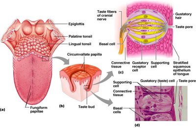
Anatomy of Taste Buds
Taste buds are the sensory organs for taste, primarily located on the tongue within papillae. There are three types of papillae: filiform, fungiform, and circumvallate. Only fungiform and circumvallate papillae contain taste buds.
Taste bud structure: Each taste bud is gourd-shaped and consists of three major epithelial cell types:
Supporting cells: Insulate the receptor cells.
Basal cells: Act as stem cells, regenerating new taste cells.
Gustatory (taste) cells: The actual receptor cells for taste.
Taste Sensations and Physiology
There are five basic taste sensations:
Sweet: Sugars, saccharin, alcohol, and some amino acids.
Salt: Metal ions (e.g., Na+, K+).
Sour: Hydrogen ions (acids).
Bitter: Alkaloids such as quinine and nicotine.
Umami: Elicited by the amino acid glutamate.
For a chemical to be tasted, it must be dissolved in saliva and contact the gustatory hairs of taste cells.
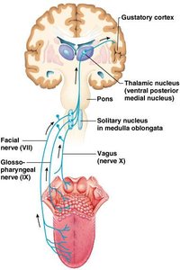
Gustatory Pathway
Taste signals are transmitted from the taste buds to the brain via cranial nerves:
Facial nerve (VII): Anterior two-thirds of the tongue.
Glossopharyngeal nerve (IX): Posterior one-third of the tongue.
Vagus nerve (X): Taste buds in the epiglottis and lower pharynx.
These nerves carry impulses to the solitary nucleus of the medulla, then to the thalamus, and finally to the gustatory cortex for perception.
Influence of Other Sensations on Taste
Taste is closely linked to smell (about 80% of taste is actually smell). Other receptors such as thermoreceptors, mechanoreceptors, and nociceptors also influence taste perception. Temperature and texture can enhance or detract from taste.
Influence of Taste on the Digestive System
Taste triggers reflexes involved in digestion, such as saliva secretion and gastric juice production, primarily via parasympathetic fibers.
It can also trigger protective reflexes like gagging.
Chemical Senses: Smell (Olfaction)
Anatomy of the Olfactory Epithelium
The organ of smell is the olfactory epithelium, located in the superior nasal concha. Olfactory receptor cells are bipolar neurons with olfactory cilia that detect odorants dissolved in mucus. Supporting cells cushion the receptors, and basal cells regenerate new olfactory neurons.
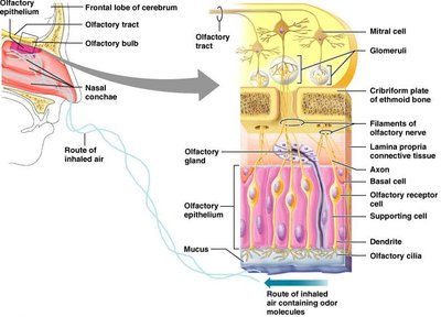
Olfactory Pathway
Olfactory receptor cells synapse with mitral cells in the olfactory bulb. Mitral cells process odor signals and send impulses to:
The olfactory cortex (for identification and interpretation)
The hypothalamus, amygdala, and limbic system (for emotional and behavioral responses)
Humans can distinguish about 10,000 different odors, and odorants must be dissolved in mucus to be detected.
Physical Senses: Sight (Vision)
Accessory Structures of the Eye
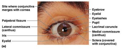
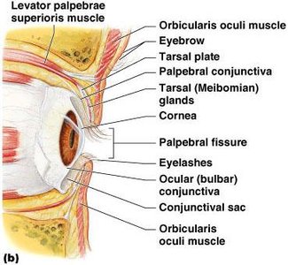
The eye is protected and supported by several accessory structures:
Eyebrows: Shade the eye and prevent sweat from entering.
Eyelids (palpebrae): Protect the eye anteriorly; contain tarsal plates for support and glands for lubrication.
Eyelashes: Initiate reflex blinking and are associated with sebaceous and ciliary glands.
Conjunctiva: Transparent mucous membrane lining the eyelids and covering the sclera, lubricating and protecting the eye.
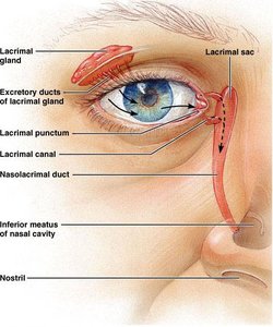
Lacrimal Apparatus
The lacrimal apparatus consists of the lacrimal gland and ducts that produce and drain tears. Tears moisten, lubricate, cleanse, and protect the eye, containing mucus, antibodies, and lysozyme.
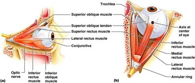
Extrinsic Eye Muscles
Six straplike extrinsic eye muscles control eye movement and maintain the shape of the eyeball. Four rectus muscles originate from the annular ring, and two oblique muscles move the eye in the vertical plane.
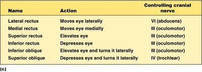
Summary Table: Extrinsic Eye Muscles and Innervation
Name | Action | Controlling cranial nerve |
|---|---|---|
Lateral rectus | Moves eye laterally | VI (abducens) |
Medial rectus | Moves eye medially | III (oculomotor) |
Superior rectus | Elevates eye | III (oculomotor) |
Inferior rectus | Depresses eye | III (oculomotor) |
Inferior oblique | Elevates eye and turns it laterally | III (oculomotor) |
Superior oblique | Depresses eye and turns it laterally | IV (trochlear) |
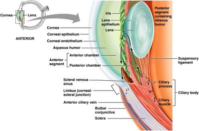
Structure of the Eyeball
The eyeball is a slightly irregular hollow sphere with anterior and posterior poles. Its wall is composed of three tunics: fibrous (sclera and cornea), vascular (choroid, ciliary body, iris), and sensory (retina). The lens separates the internal cavity into anterior and posterior segments filled with humors.
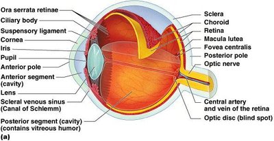
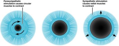
Vascular Tunic: Iris and Pupil
The iris is the colored part of the eye, with the pupil at its center. The pupil regulates the amount of light entering the eye by constricting (in bright light) or dilating (in dim light or emotional states).
Sensory Tunic: Retina
The retina consists of a pigmented layer (absorbs light) and a neural layer (contains photoreceptors, bipolar cells, and ganglion cells). The fovea centralis is the area of highest visual acuity.
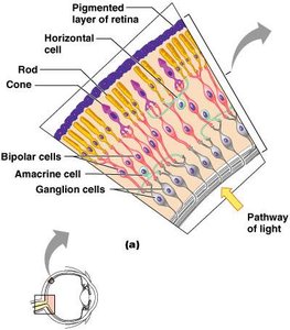
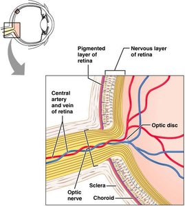
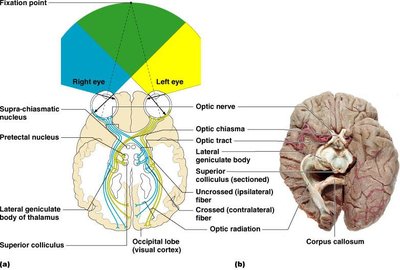
Ganglion Cells and the Optic Disc
Ganglion cell axons form the optic nerve, which exits at the optic disc (blind spot, lacking photoreceptors).
Inner Chambers and Fluids
The lens divides the eye into anterior and posterior segments. The anterior segment contains aqueous humor, while the posterior segment contains vitreous humor. These fluids maintain intraocular pressure and nourish the eye.
Visual Pathways
Axons of retinal ganglion cells form the optic nerve. Medial fibers decussate at the optic chiasm, and most fibers continue to the thalamus, then to the visual cortex. Some fibers project to the midbrain for reflexes.
Physical Senses: Hearing and Equilibrium
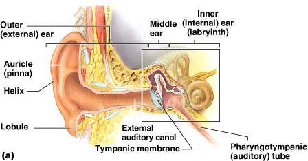
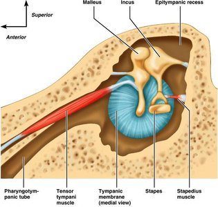
Anatomy of the Ear
The ear is divided into three parts: outer, middle, and inner ear. The outer and middle ear are involved in hearing, while the inner ear is involved in both hearing and equilibrium.
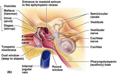
Ear Ossicles
The middle ear contains three small bones (ossicles): malleus, incus, and stapes. These transmit vibrations from the tympanic membrane to the oval window of the inner ear.
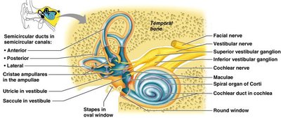
Inner Ear: Bony and Membranous Labyrinth
The inner ear consists of the bony labyrinth (filled with perilymph) and the membranous labyrinth (filled with endolymph). Major structures include the vestibule, cochlea, and semicircular canals.


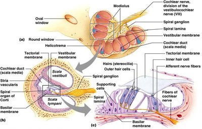
The Cochlea and Organ of Corti
The cochlea is a spiral, conical chamber containing the cochlear duct, which houses the organ of Corti (the hearing receptor). The cochlea is divided into three chambers: scala vestibuli, scala media, and scala tympani.
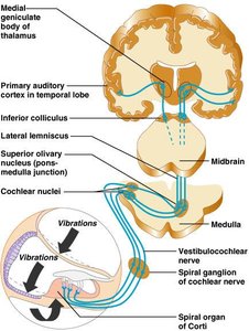
Auditory Pathway to the Brain
Sound vibrations are transmitted from the outer ear to the cochlea, where they stimulate hair cells in the organ of Corti. Impulses travel via the cochlear nerve to the cochlear nuclei, then to the thalamus, inferior colliculus, and finally the auditory cortex. Auditory pathways decussate, so both cortices receive input from both ears.
Equilibrium and Orientation
The vestibular apparatus (semicircular canals and vestibule) contains equilibrium receptors. Maculae in the vestibule monitor static equilibrium (linear acceleration), while cristae in the semicircular canals monitor dynamic equilibrium (rotational movements).
Maculae: Contain hair cells with stereocilia and a kinocilium embedded in the otolithic membrane, which contains otoliths (CaCO3 crystals).
Crista ampullaris: Located in the ampulla of each semicircular canal, responds to angular movements.
Activation of these receptors sends signals via the vestibular nerve (part of CN VIII) to the brain, allowing perception of head position and movement.
Additional info: The notes above are expanded with academic context to ensure completeness and clarity for college-level study. All images included are directly relevant to the adjacent explanations and reinforce the anatomical and physiological concepts discussed.
