 Back
BackChapter 15: The Special Senses – Structure and Function
Study Guide - Smart Notes
 Tailored notes based on your materials, expanded with key definitions, examples, and context.
The Special Senses: Overview
Introduction to Special Senses
The special senses include olfaction (smell), gustation (taste), vision, equilibrium (balance), and hearing. Unlike general senses (such as touch), these senses rely on specialized receptor cells and complex neural pathways to deliver information to the brain. Each special sense is associated with a specific organ and unique sensory receptors.
Olfaction: Sense of smell
Gustation: Sense of taste
Vision: Sense of sight
Equilibrium: Sense of balance
Hearing: Sense of sound
All special senses begin with either dendrites of specialized neurons or specialized cells that synapse with sensory neurons. A generator potential is a depolarization of the membrane that, if strong enough, triggers action potentials sent to the central nervous system (CNS).
Olfaction (Smell)
Olfactory Receptors and Pathways
Olfaction is the detection of airborne chemicals (odorants) by specialized chemoreceptive neurons in the nasal cavity. Olfactory organs are paired structures located on each side of the nasal septum and contain olfactory receptor cells distributed along the cribriform plate, superior portion of the perpendicular plate, and superior nasal conchae.
Olfactory epithelium: Contains olfactory receptor cells; odorants bind to receptors on dendrites.
Lamina propria: Areolar tissue providing support.
When odorants bind to olfactory receptors, they trigger a generator potential. If the stimulus is strong enough, action potentials are sent to the CNS. The olfactory pathway involves axons passing through the cribriform plate to the olfactory bulb, then traveling via the olfactory tract to the olfactory cortex, hypothalamus, and limbic system. This explains why smells can evoke strong emotional and behavioral responses.
Gustation (Taste)
Structure and Function of Taste Buds
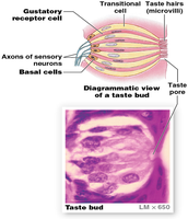
Gustation is the sense of taste, relying on chemoreceptor cells located in taste buds, primarily on the superior surface of the tongue. Taste buds are found within lingual papillae, which are epithelial projections on the tongue. There are four types of lingual papillae: vallate, foliate, fungiform, and filiform (the latter does not contain taste buds).
Taste buds: Sensory structures containing gustatory receptor cells, basal cells, and transitional cells.
Taste hairs (microvilli): Extend through a taste pore to detect dissolved chemicals.
Taste Sensations and Pathways
There are five primary taste sensations:
Sweet: Simple sugars (e.g., glucose, fructose)
Sour: Hydrogen ions (e.g., citric acid)
Salty: Metal ions (e.g., sodium, potassium)
Bitter: Various compounds, often toxic (e.g., alkaloids)
Umami: Amino acids, especially glutamate (savory)
Water receptors are also present, mainly in the pharynx, and influence water balance and blood volume via the hypothalamus.
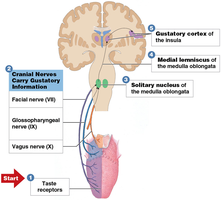
Neural Pathways for Taste
Taste information is carried by the facial (VII), glossopharyngeal (IX), and vagus (X) nerves. These axons terminate in the solitary nucleus of the medulla oblongata, then relay to the thalamus and finally to the primary gustatory cortex in the parietal lobe. Integration with visual and olfactory stimuli occurs in the insula and inferior frontal lobe, contributing to the overall perception of flavor and emotional responses to taste.
Vision
Accessory Structures of the Eye
The eye is protected and maintained by several accessory structures:
Eyelashes: Prevent foreign matter from entering the eye.
Eyelids (palpebrae): Protect and lubricate the eye through blinking.
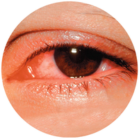
Conjunctiva: Mucous membrane lining eyelids and covering the anterior eye; inflammation is called conjunctivitis (pinkeye).
Lacrimal apparatus: Produces and drains tears, which lubricate, nourish, and protect the eye.
Structure of the Eyeball
The eyeball consists of three layers (tunics):
Fibrous layer: Sclera and cornea
Vascular layer: Iris, choroid, ciliary body
Inner layer: Retina
The eye contains two main cavities:
Anterior cavity: Contains aqueous humor; divided into anterior and posterior chambers
Posterior cavity: Contains vitreous body (gelatinous mass with vitreous humor)
Retinal Structure and Photoreceptors
The retina contains photoreceptors (rods and cones), pigment cells, supporting cells, and neurons. The macula is a region with a high density of photoreceptors, and the fovea centralis is the point of sharpest vision.
Rods: Highly sensitive, enable vision in dim light, provide black-and-white vision
Cones: Responsible for color vision (blue, green, red cones), require more light, provide sharper images
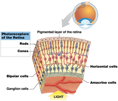
Rods and cones synapse with bipolar cells, which in turn synapse with ganglion cells. Visual pigments in photoreceptors (derived from rhodopsin) transduce light into neural signals.
Visual Pathways and Refractive Problems
Visual information travels from the retina through the optic nerves, optic chiasm, and optic tracts to the lateral geniculate nucleus, midbrain, and primary visual cortex. Refractive problems include:
Emmetropia: Normal vision
Myopia: Nearsightedness; image focuses in front of retina, corrected with concave lenses
Hyperopia: Farsightedness; image focuses beyond retina, corrected with convex lenses
Surgical corrections include PRK and LASIK, which reshape the cornea.
Equilibrium and Hearing
Anatomy of the Ear
The ear is divided into three regions:
External ear: Collects sound waves (auricle, external acoustic meatus, ceruminous glands)
Middle ear: Air-filled chamber with tympanic membrane and auditory ossicles (malleus, incus, stapes)
Internal ear: Contains sensory organs for hearing and equilibrium, protected by the bony labyrinth
Internal Ear and Sensory Receptors
The bony labyrinth surrounds the membranous labyrinth and is filled with perilymph. The internal ear consists of:
Semicircular canals: Detect rotational movements (equilibrium)
Vestibule (utricle and saccule): Detect gravity and linear acceleration
Cochlea: Contains the cochlear duct and spiral organ (receptors for hearing)
Hair cells are mechanoreceptors that transduce mechanical stimuli (sound waves or head movement) into electrical signals.
Hearing Pathways
Sound waves move the tympanic membrane, which transmits vibrations through the auditory ossicles to the oval window of the inner ear. This causes movement of fluid in the cochlea, stimulating hair cells. The neural pathway for hearing involves:
Depolarization of hair cells triggers action potentials in the cochlear nerve
Axons synapse in the pons, where sound localization occurs
Signals are relayed to the midbrain (startle reflex), thalamus (sound processing), and primary auditory cortex
Most auditory information from one cochlea is projected to the opposite side of the brain, aiding in sound localization and reducing the impact of unilateral damage.
Disorders of the Special Senses
Vision Disorders
Senile cataract: Lens loses transparency with age; can lead to blindness but is surgically correctable
Presbyopia: Age-related loss of lens elasticity, causing difficulty focusing on close objects
Equilibrium Disorders
Vertigo: False sensation of spinning, often due to inner ear or vestibular nerve dysfunction
Motion sickness: Most common cause of vertigo; symptoms include headache, nausea, and vomiting
Hearing Disorders
Conductive hearing loss: Impaired conduction of sound waves (e.g., earwax, infection, perforated eardrum)
Sensorineural hearing loss: Damage to cochlea or neural pathways (e.g., loud noise, trauma, aging)
Age-related changes: Stiffening of tympanic membrane and ossicles, ossification of round window
