 Back
BackChapter 15: The Special Senses – Structure and Function
Study Guide - Smart Notes
 Tailored notes based on your materials, expanded with key definitions, examples, and context.
The Special Senses
Introduction
The special senses include smell (olfaction), taste (gustation), vision, hearing, and equilibrium. These senses rely on specialized sensory receptors located in distinct organs, allowing the body to detect and interpret complex environmental stimuli.
Smell (Olfaction)
Anatomy of the Olfactory System
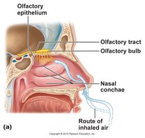
The olfactory receptors are located in the olfactory epithelium, which covers the cribriform plate and the superior nasal concha. Inhaled air carries odorant molecules to this region, where they interact with sensory neurons.
Olfactory epithelium: Contains olfactory receptor cells, supporting cells, and basal cells (stem cells for regeneration).
Olfactory bulb: The site where olfactory neuron axons synapse before signals are relayed to the brain via the olfactory tract.
Olfactory tract: Carries olfactory information to the olfactory cortex and limbic system for processing.
Olfactory Receptors and Signal Transduction
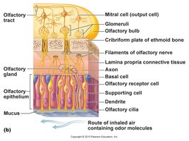
Olfactory receptor cells are bipolar neurons with cilia that extend into the mucus layer. Odorant molecules dissolve in the mucus and bind to receptors on these cilia, initiating a signal transduction cascade.
Supporting cells: Insulate olfactory cells and help move mucus with their cilia.
Basal cells: Stem cells that regenerate olfactory neurons.
Signal pathway: Odorant binds receptor → axons pass through cribriform plate → synapse in olfactory bulb → olfactory tract → olfactory cortex/limbic system.
Taste (Gustation)
Anatomy of Taste Buds and Papillae
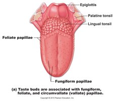
Taste buds are the primary receptors for gustation and are located on the tongue and oral cavity. The tongue contains three types of papillae, two of which (fungiform and circumvallate/foliate) contain taste buds in adults.
Taste buds: Found on fungiform, foliate, and circumvallate papillae.
Function: Taste triggers reflexes that prepare the GI tract for digestion and can induce vomiting if harmful substances are detected.
Structure of a Taste Bud
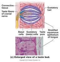
Each taste bud consists of gustatory (taste) cells, basal cells (stem cells), and supporting cells. Gustatory hairs project through a taste pore and interact with dissolved chemicals (tastants).
Gustatory cells: Sensory cells that synapse with cranial nerve fibers.
Basal cells: Regenerate gustatory cells.
Signal pathway: Tastant binds receptor → gustatory cell transmits signal to cranial nerves VII, IX, X → medulla → thalamus → gustatory cortex/limbic system.
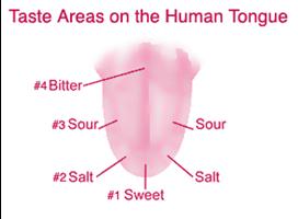
Primary Taste Sensations
There are five primary taste sensations: sweet, sour, salty, bitter, and umami (savory). Each is detected by specific receptors distributed across the tongue, though all areas can detect all tastes to some degree.
Sweet: Indicates energy-rich nutrients.
Sour: Detects acidity.
Salty: Senses sodium ions.
Bitter: Often signals potential toxins.
Umami: Detects amino acids (e.g., glutamate).
Vision
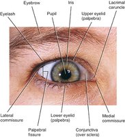
Accessory Structures of the Eye
The eye is protected and supported by several accessory structures, including the eyelids, eyelashes, conjunctiva, and lacrimal apparatus.
Eyelids: Protect the eye, spread lubricant, and anchor muscles.
Eyelashes: Trigger the blink reflex.
Conjunctiva: Thin mucous membrane lining the eyelids and covering the sclera for protection.
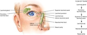
Lacrimal apparatus: Produces tears containing mucus, antibodies, and lysozyme for lubrication and protection.
Structure of the Eyeball
The eyeball is a hollow sphere with anterior and posterior poles, divided by the lens into anterior and posterior cavities. The walls of the eyeball have three layers (tunicas): fibrous, vascular, and sensory.
Fibrous tunica: Outer layer; includes the sclera (white, maintains shape) and cornea (transparent, regenerates quickly).
Vascular tunica (uvea): Middle layer; includes the choroid (blood supply, pigment), ciliary body (muscle and aqueous humor production), and iris (controls pupil size).
Sensory tunica (retina): Inner layer; contains photoreceptors (rods and cones) and association neurons.
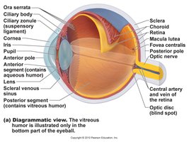
Lens and Humors
The lens focuses light on the retina and is adjustable by ciliary muscles. The anterior cavity contains aqueous humor (nourishes tissues, maintains pressure), while the posterior cavity contains vitreous humor (maintains shape, never replaced).
Lens: Biconvex, transparent, flexible, avascular; contains crystallin protein.
Cataracts: Clouding of the lens, leading to vision impairment.
Glaucoma: Increased intraocular pressure due to blocked aqueous humor drainage.
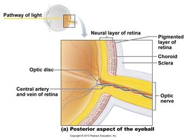
Retina and Photoreceptors
The retina contains two layers: a pigmented layer (absorbs light, stores vitamin A) and a neural layer (contains rods and cones). The macula lutea and fovea centralis are regions of sharpest vision, while the optic disc is a blind spot lacking photoreceptors.
Rods: Sensitive to low light, responsible for night and peripheral vision, perceive only gray tones.
Cones: Require bright light, responsible for color vision, concentrated in the fovea centralis.

Pathway of Light and Visual Signal Transmission
Light passes through the cornea, aqueous humor, lens, and vitreous humor before reaching the photoreceptors in the retina. Neural signals are then transmitted through bipolar and ganglion cells, forming the optic nerve, which carries information to the brain.
Pathway: Cornea → aqueous humor → lens → vitreous humor → retina (photoreceptors) → bipolar cells → ganglion cells → optic nerve → brain.
Hearing and Equilibrium
Anatomy of the Ear
The ear is divided into three regions: external, middle, and inner ear. Each region plays a role in hearing and balance.
External ear: Collects sound waves and directs them to the tympanic membrane (eardrum).
Middle ear: Air-filled cavity containing ossicles (malleus, incus, stapes) that transmit vibrations to the inner ear.
Inner ear: Contains the vestibule, semicircular canals (equilibrium), and cochlea (hearing).
Physiology of Hearing
Sound waves cause movement of the basilar membrane in the cochlea, bending hair cells against the tectorial membrane. This stimulates dendrites of the cochlear nerve, sending signals to the brain.
Intensity (loudness): Coded by the degree of hair cell deflection.
Frequency (pitch): Coded by the location of deflection on the basilar membrane.
Equilibrium
The vestibule and semicircular canals of the inner ear contain receptors for balance. The utricle and saccule detect linear acceleration, while the semicircular canals detect rotational movements.
Utricle: Detects forward and backward motion.
Saccule: Detects up and down motion.
Semicircular canals: Detect rotational movements in three planes.
Clinical Considerations
Vision Impairments
Myopia (nearsightedness): Focal point is in front of the retina; corrected with concave lenses.
Hyperopia (farsightedness): Focal point is behind the retina; corrected with convex lenses.
Color blindness: Inability to distinguish certain colors due to absent or nonfunctional cones.
Macular degeneration: Loss of central vision due to degeneration of the macula lutea.
Hearing Loss
Conductive deafness: Impaired conduction of sound waves through the external or middle ear.
Sensorineural deafness: Damage to inner ear structures or auditory nerve.
Central deafness: Damage to neural pathways in the brain.
Equilibrium Disorders
Motion sickness: Occurs when visual information conflicts with vestibular input, leading to nausea and vomiting.
