 Back
BackChapter 15: The Special Senses – Vision, Smell, Taste, Hearing, and Equilibrium
Study Guide - Smart Notes
 Tailored notes based on your materials, expanded with key definitions, examples, and context.
The Special Senses
Overview of Special Senses
The special senses include vision, taste, smell, hearing, and equilibrium. Unlike general senses, which are mediated by simple receptors distributed throughout the body, special senses rely on complex, localized sensory organs primarily in the head. Each sense uses specialized receptor cells to detect specific stimuli and transmit information to the brain for interpretation.
The Eye and Vision
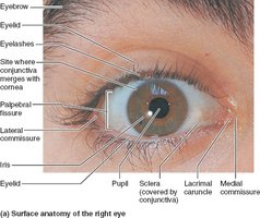
Accessory Structures of the Eye
The accessory structures of the eye protect and support its function. These include the eyebrows, eyelids, conjunctiva, lacrimal apparatus, and extrinsic eye muscles.
Eyebrows: Shade the eyes and prevent sweat from reaching them.
Eyelids (palpebrae): Protect the eye anteriorly, contain glands for lubrication, and blink reflexively to moisten and shield the eye.
Conjunctiva: Transparent mucous membrane lining the eyelids and covering the sclera, producing lubricating mucus.
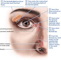
Lacrimal apparatus: Produces and drains tears, which contain mucus, antibodies, and lysozyme for eye protection.
Extrinsic eye muscles: Six muscles control eye movement and maintain its shape.

Lacrimal Apparatus
The lacrimal apparatus consists of the lacrimal gland and ducts that drain tears into the nasal cavity. Tears lubricate, cleanse, and protect the eye surface.
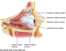
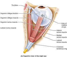
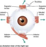
Extrinsic Eye Muscles
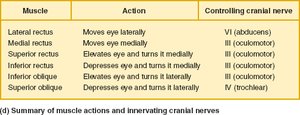
Six extrinsic muscles move the eyeball. Four are rectus muscles (superior, inferior, lateral, medial), and two are oblique (superior, inferior). These muscles allow precise control of eye movement and are innervated by cranial nerves III, IV, and VI.
Muscle | Action | Controlling Cranial Nerve |
|---|---|---|
Lateral rectus | Moves eye laterally | VI (abducens) |
Medial rectus | Moves eye medially | III (oculomotor) |
Superior rectus | Elevates eye, turns it medially | III (oculomotor) |
Inferior rectus | Depresses eye, turns it medially | III (oculomotor) |
Inferior oblique | Elevates eye, turns it laterally | III (oculomotor) |
Superior oblique | Depresses eye, turns it laterally | IV (trochlear) |
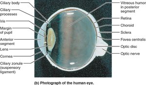
Structure of the Eyeball
The eyeball is a nearly spherical organ with three layers: fibrous, vascular, and inner (retina). It contains the lens and is divided into anterior and posterior segments by the lens.
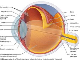
Fibrous Layer
Sclera: Opaque, white, protective outer layer; continuous with the dura mater of the brain.
Cornea: Transparent anterior part; allows light entry and bends light for focusing.
Vascular Layer (Uvea)
Choroid: Pigmented, vascular layer supplying blood and absorbing light.
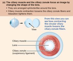
Ciliary body: Contains ciliary muscles (control lens shape), ciliary processes (secrete aqueous humor), and ciliary zonule (holds lens).
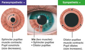
Iris: Colored part; controls pupil size to regulate light entry.
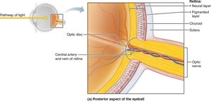
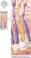
Inner Layer (Retina)
Pigmented layer: Absorbs light, stores vitamin A, and phagocytizes debris.
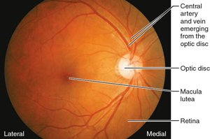
Neural layer: Contains photoreceptors (rods and cones), bipolar cells, and ganglion cells. The optic disc is the blind spot where the optic nerve exits.
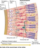
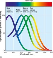
Photoreceptors
Rods: Sensitive to dim light, provide peripheral and night vision, but no color.
Cones: Detect bright light and color, concentrated in the fovea centralis for sharp vision.
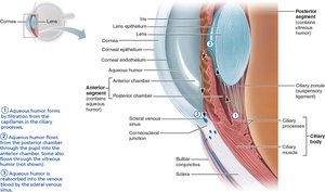
Internal Chambers and Fluids
Vitreous humor: Gel-like, fills posterior segment, supports retina, transmits light.
Aqueous humor: Watery, fills anterior segment, nourishes lens and cornea, drains via scleral venous sinus.
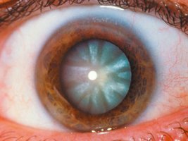
Lens
The lens is a biconvex, transparent, flexible structure that focuses light on the retina. With age, it becomes denser and less elastic, leading to presbyopia and increased risk of cataracts.
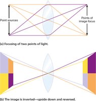
Cornea and Lens Focus Light on the Retina
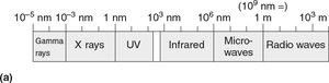
Wavelength and Color
Visible light is a small part of the electromagnetic spectrum (400–700 nm). The color perceived depends on the wavelength reflected by objects.
Refraction and Lenses
Refraction is the bending of light as it passes through different media. The cornea and lens refract light to focus it on the retina. Convex lenses converge light rays; concave lenses diverge them.

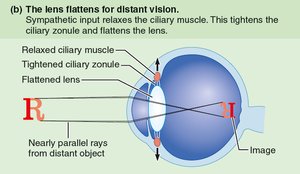
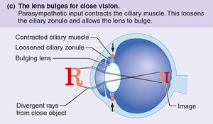
Focusing for Distant and Close Vision
Distant vision: Lens is flat; ciliary muscles relaxed.
Close vision: Lens bulges; ciliary muscles contract. Requires accommodation, pupil constriction, and convergence of the eyeballs.
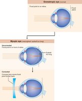
Problems of Refraction
Myopia (nearsightedness): Eyeball too long; corrected with concave lenses.
Hyperopia (farsightedness): Eyeball too short; corrected with convex lenses.
Astigmatism: Unequal curvature; corrected with cylindrical lenses or surgery.

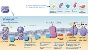
Phototransduction
Photoreceptor Structure and Function
Photoreceptors (rods and cones) convert light into electrical signals. Each has an outer segment (with visual pigments) and an inner segment (with cell body and synaptic terminal).
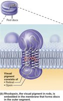
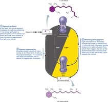
Visual Pigments and Phototransduction
Rhodopsin: Visual pigment in rods, formed from opsin and 11-cis-retinal (from vitamin A).
Light converts 11-cis-retinal to all-trans-retinal, triggering a cascade that hyperpolarizes the cell and initiates vision.
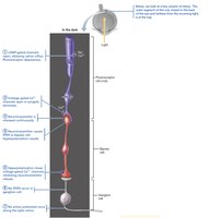
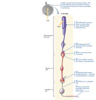
Signal Transmission in the Retina
Light hyperpolarizes photoreceptors, reducing glutamate release. This excites bipolar cells, which stimulate ganglion cells to generate action potentials sent to the brain.
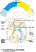
Visual Pathways and Processing
Visual Pathway to the Brain
Axons of ganglion cells form the optic nerve, which partially crosses at the optic chiasma. Visual information is relayed to the thalamus and then to the primary visual cortex for interpretation.
The Chemical Senses: Smell and Taste
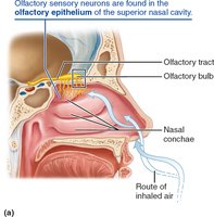
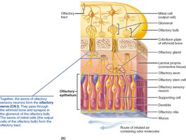
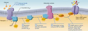
Olfaction (Smell)
Olfactory receptors are located in the nasal cavity and detect volatile chemicals dissolved in mucus. Each receptor responds to specific odorants, and the olfactory pathway transmits signals to the olfactory cortex and limbic system.
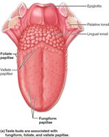
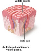
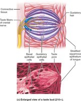
Taste (Gustation)
Taste buds, mainly on the tongue, detect five basic tastes: sweet, sour, salty, bitter, and umami. Taste transduction involves chemical binding, cell depolarization, and neurotransmitter release to sensory neurons.
The Ear: Hearing and Equilibrium
Ear Structure and Function
The ear is divided into external, middle, and internal regions. The external and middle ear are involved in hearing, while the internal ear is responsible for both hearing and equilibrium.
External ear: Auricle and external acoustic meatus funnel sound to the tympanic membrane.
Middle ear: Contains auditory ossicles (malleus, incus, stapes) that transmit vibrations to the oval window.
Internal ear: Houses the cochlea (hearing) and vestibular apparatus (equilibrium).
*Additional info: For a complete understanding of hearing and equilibrium, refer to diagrams of the cochlea, auditory pathway, and vestibular system as described in the text.*
