 Back
BackChapter 16: The Autonomic Nervous System and Higher-Order Functions – Study Notes
Study Guide - Smart Notes
 Tailored notes based on your materials, expanded with key definitions, examples, and context.
The Autonomic Nervous System (ANS) and Higher-Order Functions
Introduction to the ANS and Higher-Order Functions
The autonomic nervous system (ANS), also known as the visceral motor system, operates without conscious control and regulates the activity of visceral effectors such as smooth muscle, glands, cardiac muscle, and adipocytes. It is essential for coordinating cardiovascular, respiratory, digestive, urinary, and reproductive functions. Higher-order functions include consciousness, learning, and intelligence, all of which require the cerebral cortex and involve both conscious and unconscious information processing.
Organization of the Autonomic Nervous System
Comparison with the Somatic Nervous System (SNS)
The somatic nervous system (SNS) controls voluntary movements of skeletal muscles, while the ANS controls involuntary actions of visceral effectors. The hypothalamus contains integrative centers for the ANS, and motor neurons of the CNS synapse on visceral motor neurons in autonomic ganglia.
Somatic Nervous System: Voluntary, single motor neuron pathway, targets skeletal muscle.
Autonomic Nervous System: Involuntary, two-neuron pathway (preganglionic and postganglionic), targets smooth muscle, cardiac muscle, glands, and adipocytes.
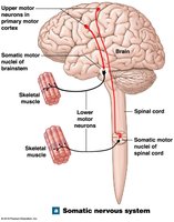
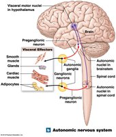
Divisions of the ANS
Sympathetic Division: Prepares the body for emergencies ("fight or flight"), increases alertness, metabolic rate, and muscular abilities.
Parasympathetic Division: Conserves energy and maintains resting metabolic rate ("rest and digest").
Functional Interactions
Sympathetic and parasympathetic divisions often have opposing effects but may also work independently or together in complex processes.
Sympathetic Division (Thoracolumbar Division)
Structure and Pathways
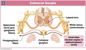
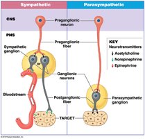
The sympathetic division has short preganglionic fibers originating from the thoracic and lumbar segments (T1–L2) and long postganglionic fibers. Ganglionic neurons are located in ganglia near the spinal cord.
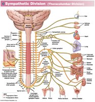
Sympathetic Chain Ganglia: Located on either side of the vertebral column, control effectors in the body wall, thoracic cavity, head, neck, and limbs.
Collateral Ganglia: Anterior to vertebral bodies, innervate abdominopelvic tissues and viscera.
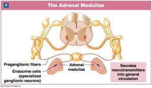
Adrenal Medullae: Modified sympathetic ganglia that release neurotransmitters (epinephrine and norepinephrine) into the bloodstream.
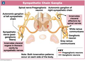
Sympathetic Responses
Increased alertness and metabolic rate
Reduced digestive and urinary functions
Activation of energy reserves, increased heart rate, blood pressure, and respiratory rate
Activation of sweat glands
Neurotransmitters and Receptors
Preganglionic neurons release acetylcholine (ACh) (always excitatory).
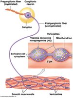
Most postganglionic neurons release norepinephrine (NE) (adrenergic), some release ACh (cholinergic), and a few release nitric oxide (NO).
Adrenergic receptors: Alpha (α) and Beta (β) receptors, both G-protein-coupled.
Alpha-1: Excitatory (smooth muscle contraction); Alpha-2: Inhibitory (lowers cAMP).
Beta-1: Increases metabolic activity; Beta-2: Relaxes smooth muscle; Beta-3: Lipolysis in adipocytes.
Parasympathetic Division (Craniosacral Division)
Structure and Pathways
The parasympathetic division has long preganglionic fibers from the brainstem and sacral spinal cord (S2–S4), and short postganglionic fibers located in or near target organs.
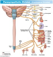
Terminal Ganglia: Near target organs, usually paired.
Intramural Ganglia: Embedded within target organ tissues.
Major outflow via the vagus nerve (CN X), which provides 75% of all parasympathetic outflow.
Parasympathetic Responses
Decreased metabolic rate, heart rate, and blood pressure
Increased secretion by digestive glands and motility in the digestive tract
Stimulation of urination and defecation
Constriction of pupils and respiratory passageways
Neurotransmitters and Receptors
All parasympathetic neurons release ACh.
Nicotinic receptors: On ganglion cells, always excitatory.
Muscarinic receptors: On effector cells, can be excitatory or inhibitory, longer-lasting effects.
Comparison of Sympathetic and Parasympathetic Divisions
The sympathetic division has widespread effects, extensive divergence, and uses both ACh and NE. The parasympathetic division has more specific, localized effects, less divergence, and uses only ACh.
Dual Innervation and Autonomic Tone
Dual Innervation
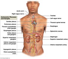
Most vital organs receive input from both ANS divisions, often with opposing effects. Autonomic plexuses are networks of sympathetic and parasympathetic fibers that innervate organs.
Examples: Cardiac, pulmonary, esophageal, celiac, and hypogastric plexuses.
Autonomic Tone
Autonomic motor neurons maintain a resting level of activity, allowing for increases or decreases in function.
Example: The heart receives both sympathetic (increases rate) and parasympathetic (decreases rate) input.
Some organs, like blood vessels, are innervated by only one division (sympathetic).
Regulation of Autonomic Functions
Visceral Reflexes
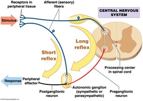
Visceral reflexes are automatic, polysynaptic reflexes that control visceral effectors. They can be long (involving the CNS) or short (bypassing the CNS, occurring in autonomic ganglia).
Long reflexes: Coordinate activities of entire organs.
Short reflexes: Control localized responses, especially in the digestive tract (enteric nervous system).
Hierarchy of Control
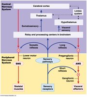
Centers in the medulla oblongata and hypothalamus regulate complex autonomic functions.
Integration of ANS and SNS occurs at the brainstem level.
Higher-Order Functions
Memory and Learning
Higher-order functions require the cerebral cortex and involve both conscious and unconscious processing. Memories are classified as fact (specific information) or skill (learned motor behaviors). Memory consolidation is the process of converting short-term to long-term memory.
Short-term memory: Temporary, easily lost unless consolidated.
Long-term memory: Secondary (fade with time) and tertiary (permanent).
Key brain regions: Amygdaloid body, hippocampus, nucleus basalis, cerebral cortex.
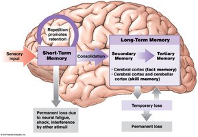
Cellular Mechanisms of Memory
Increased neurotransmitter release, facilitation at synapses, and formation of additional synaptic connections underlie memory formation.
Memory engrams are neural circuits formed by experience and repetition.
NMDA receptors in the hippocampus are crucial for long-term memory formation.
States of Consciousness and Sleep
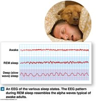
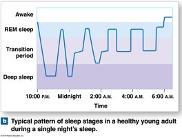
Deep sleep (NREM): Body relaxes, reduced physiological activity.
REM sleep: Active dreaming, EEG resembles awake state, muscle tone decreases.
Sleep alternates between REM and deep sleep; lack of sleep impairs mental function.
Arousal and the Reticular Activating System (RAS)
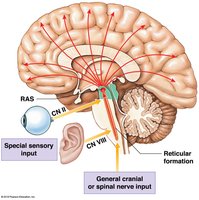
Arousal is mediated by the reticular formation and RAS, which project to the cerebral cortex.
Stimulation of RAS produces widespread cortical activation; sleep is ended by RAS activation.
Neurotransmitters such as NE and serotonin regulate sleep-wake cycles.
Neurotransmitters and Brain Function
Serotonin: Affects mood and sensory interpretation; imbalances can cause depression or hallucinations.
Dopamine: Involved in motor control and reward; imbalances linked to Parkinson’s disease and schizophrenia.
Effects of Aging on the Nervous System
Anatomical and Functional Changes
Reduction in brain size and weight, especially in the cerebral cortex.
Decrease in neuron number, synaptic connections, and neurotransmitter production.
Accumulation of intracellular deposits (lipofuscin, neurofibrillary tangles) and extracellular plaques (amyloid protein).
Functional changes include memory loss, slower reaction times, and reduced sensory acuity.
Senile dementia (e.g., Alzheimer’s disease) may develop in some elderly individuals.
Summary Table: Sympathetic vs. Parasympathetic Divisions
Feature | Sympathetic Division | Parasympathetic Division |
|---|---|---|
Origin | Thoracolumbar (T1–L2) | Craniosacral (Brainstem, S2–S4) |
Preganglionic Fiber Length | Short | Long |
Postganglionic Fiber Length | Long | Short |
Main Neurotransmitter (Postganglionic) | Norepinephrine (NE) | Acetylcholine (ACh) |
General Function | "Fight or flight" | "Rest and digest" |
