 Back
Chapter 16: The Autonomic Nervous System and Higher-Order Functions – Study Notes
Study Guide - Smart Notes
 Tailored notes based on your materials, expanded with key definitions, examples, and context.
Autonomic Nervous System (ANS) Overview
Organization and Comparison with Somatic Nervous System
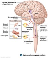
The autonomic nervous system (ANS) is responsible for involuntary control of visceral effectors, such as smooth muscle, glands, cardiac muscle, and adipocytes. It coordinates cardiovascular, respiratory, digestive, urinary, and reproductive functions. The integrative centers are located in the hypothalamus, and autonomic ganglia are clusters of cell bodies of visceral motor neurons outside the CNS.
ANS: Involuntary control; CNS visceral motor neurons synapse with visceral motor neurons in autonomic ganglia.
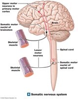
Somatic Nervous System (SNS): Voluntary control; CNS motor neurons synapse directly with skeletal muscle cells.
Visceral Motor Neurons
Preganglionic neurons: Cell bodies in brainstem and spinal cord; axons (preganglionic fibers) project to autonomic ganglia.
Postganglionic neurons: Cell bodies in autonomic ganglia; axons (postganglionic fibers) synapse with peripheral target organs.
Divisions of the ANS
Sympathetic division: "Fight or flight"; prepares the body for stress or emergency, increases alertness, respiratory rate, metabolic rate, and muscular abilities.
Parasympathetic division: "Rest and digest"; conserves energy and maintains resting metabolic rate.
The Sympathetic Division
Organization and Structure
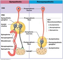
The sympathetic division is also known as the thoracolumbar division. Preganglionic neuron cell bodies are located in the lateral horns of the thoracic and lumbar spinal cord. Preganglionic fibers are short and synapse with many postganglionic neurons, releasing acetylcholine (ACh). Postganglionic neuron cell bodies are in ganglia near the spinal cord, and their fibers are long, releasing norepinephrine at peripheral synapses.
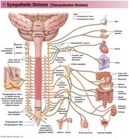
Sympathetic preganglionic fibers: Myelinated; pass through spinal anterior roots and white rami communicantes to reach sympathetic ganglia.
Types of ganglia: Sympathetic chain ganglia, collateral ganglia, adrenal medullae.
Sympathetic Chain Ganglia
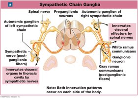
Located on either side of the vertebral column, these ganglia innervate effectors in the body wall, thoracic cavity, head, neck, and limbs. One preganglionic fiber can synapse with many postganglionic neurons. Postganglionic fibers are unmyelinated and pass through gray rami communicantes.
Collateral Ganglia
Collateral ganglia are anterior to the vertebral column and innervate abdominopelvic tissues and viscera. They reduce blood flow and energy use by organs not important for immediate survival and cause the release of stored energy.
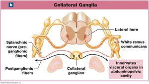
Celiac ganglia: Innervate stomach, liver, gallbladder, pancreas, spleen.
Superior mesenteric ganglion: Innervate small intestine and proximal large intestine.
Inferior mesenteric ganglion: Innervate kidneys, urinary bladder, distal large intestine, sex organs.
Adrenal Medullae
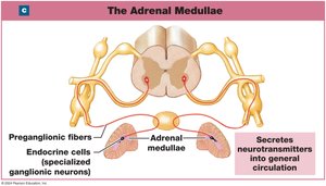
The adrenal medulla is a modified sympathetic ganglion at the center of each adrenal gland. When stimulated, it releases epinephrine and norepinephrine into the bloodstream, functioning as hormones and causing widespread metabolic changes.
Sympathetic Activation
Increased alertness, energy, blood pressure, heart rate, breathing rate, and muscle tone.
Mobilization of energy reserves.
Sympathetic Neurotransmitters and Receptors
Neurotransmitters
Preganglionic neurons: Release acetylcholine (ACh) at ganglia (cholinergic, always excitatory).
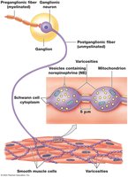
Postganglionic neurons: Release norepinephrine (NE) at target organs (adrenergic); NE can be reabsorbed or broken down by monoamine oxidase (MAO) or catechol-O-methyltransferase (COMT).
Some postganglionic neurons release ACh (body wall, skin, brain, skeletal muscle).
Adrenal medulla releases epinephrine and norepinephrine as hormones.
Receptors
Adrenergic receptors: G-protein-coupled receptors for NE and epinephrine.
Alpha receptors: More sensitive to NE; alpha-1 (excitatory, smooth muscle), alpha-2 (inhibitory, lowers cAMP).
Beta receptors: Beta-1 (increase metabolic activity), beta-2 (relaxation of smooth muscle in respiratory tract), beta-3 (lipolysis in adipocytes).
Sympathetic Synapses at Target Organs
Most are adrenergic.
Some are cholinergic (sweat glands, blood vessels of skeletal muscles and brain).
Some are nitroxidergic (release nitric oxide, cause vasodilation).
The Parasympathetic Division
Organization and Structure
The parasympathetic division is also known as the craniosacral division. Preganglionic neuron cell bodies are in the brainstem and sacral spinal cord. Preganglionic fibers are long and synapse with a few postganglionic neurons, releasing ACh. Postganglionic neuron cell bodies are in ganglia within or adjacent to target organs, and their fibers are short, also releasing ACh.
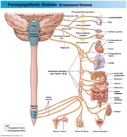
Preganglionic fibers leave the brain in cranial nerves and synapse in ciliary, pterygopalatine, submandibular, and otic ganglia.
The vagus nerve provides 75% of all parasympathetic output.
Fibers from sacral segments form pelvic nerves and synapse at intramural ganglia in the walls of organs.
Ganglia
Terminal ganglion: Near the target organ, usually paired.
Intramural ganglion: Embedded in the tissues of the target organ, consists of interconnected masses and clusters of ganglion cells.
Major Effects of Parasympathetic Activation
Constriction of pupils, focusing lenses for near vision.
Secretion by digestive glands and hormones for nutrient absorption.
Changes in blood flow and glandular activity associated with sexual arousal.
Increased smooth muscle activity in digestive tract.
Stimulation of urination and defecation.
Constriction of respiratory passageways.
Reduction in heart rate and force of contraction.
Parasympathetic Neurotransmitters and Receptors
Neurotransmitters
All parasympathetic neurons release ACh.
Most ACh is inactivated at the synapse by acetylcholinesterase (AChE); ACh that diffuses is inactivated by tissue cholinesterase.
Receptors
Cholinergic receptors: Receptors for ACh.
Nicotinic receptors: On postganglionic neurons of both divisions and at neuromuscular junctions; chemically gated Na+ channels; activation causes excitation.
Muscarinic receptors: At target organs and tissues of the parasympathetic division; G protein-coupled; response can be excitatory or inhibitory.
Environmental Toxins
Nicotine: Binds to nicotinic receptors; widespread autonomic activation, can cause coma or death.
Muscarine: Binds to muscarinic receptors; produced by some poisonous mushrooms; causes exaggerated parasympathetic effects.
Summary Comparison of ANS Divisions
Sympathetic vs. Parasympathetic
Sympathetic: Widespread effects, extensive divergence, short preganglionic/long postganglionic fibers, most postganglionic fibers release NE, receptors are adrenergic and some muscarinic cholinergic.
Parasympathetic: More specific effects, less divergence, long preganglionic/short postganglionic fibers, all neurons are cholinergic, receptors are nicotinic and muscarinic cholinergic.
Dual Innervation and Autonomic Tone
Functional Significance
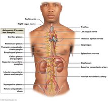
Most vital organs are innervated by both divisions, usually with opposing effects. Autonomic plexuses are nerve networks formed by mingled sympathetic and parasympathetic fibers, traveling with blood and lymphatic vessels.
Cardiac plexus: Innervates the heart.
Pulmonary plexus: Innervates the lungs.
Esophageal plexus: Innervates the esophagus.
Celiac plexus: Innervates abdominal viscera.
Inferior mesenteric plexus: Innervates abdominal organs.
Hypogastric plexus: Innervates pelvic organs.
Autonomic Tone
Resting level of activity of autonomic motor neurons.
Nerves maintain background activity, can increase or decrease activity for greater control.
Significant where dual innervation occurs, but more important where only one division innervates.
Examples
Heart: ACh slows heart rate (parasympathetic), NE accelerates heart rate (sympathetic).
Blood vessels: Only sympathetic control; NE keeps muscles partially contracted, decreased NE causes dilation.
Regulation of Autonomic Functions
Visceral Reflexes
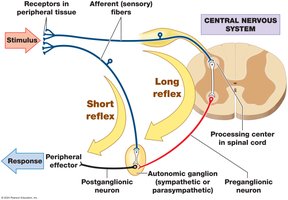
Visceral reflexes are autonomic polysynaptic reflexes providing automatic motor responses in glands and nonskeletal muscle organs. They can be modified by higher centers, especially the hypothalamus.
Reflex arc: Receptor, sensory neuron, processing center, two visceral motor neurons, peripheral effector.
Long reflexes: Coordinate activities of entire organs; sensory neurons deliver information to CNS.
Short reflexes: Bypass CNS; control activity in one part of an organ; important for digestive tract.
Enteric Nervous System
Controls digestive functions independent of CNS.
Ganglia in digestive tract walls contain cell bodies of visceral sensory neurons, interneurons, and motor neurons.
Higher Levels of Autonomic Control
Medulla oblongata coordinates complex reflexes (salivation, swallowing, digestive secretions, movement, urinary function).
Regulated by hypothalamus.
Integration of ANS and SNS Activities
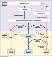
Parallels in organization and function.
Integration occurs at the brainstem; both systems are under control of higher centers.
Higher-Order Functions
Memory Creation, Storage, and Recall
Higher-order functions require the cerebral cortex, involve conscious and unconscious processing, and are subject to adjustment over time. Memories are stored information gathered through experience.
Fact memories: Specific bits of factual information.
Skill memories: Learned motor behaviors, often unconscious with repetition.
Short-term memories: Do not last long, can be recalled immediately.
Memory consolidation: Conversion of short-term to long-term memory; involves repetition.
Long-term memories: Secondary (fade with time), tertiary (last a lifetime).
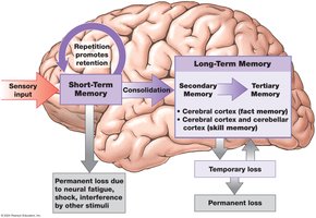
Brain Regions Involved in Memory
Amygdaloid body and hippocampus: Essential for memory consolidation; damage prevents new long-term memories.
Nucleus basalis: Role in memory storage and retrieval; damage affects emotional states and intellectual functions.
Cerebral cortex: Stores most long-term memories; association areas for sensory/motor memories.
Cellular Mechanisms of Memory Formation
Memory consolidation involves anatomical and physiological changes in neurons and synapses.
Increased neurotransmitter release, facilitation at synapses, formation of new synaptic connections.
Memory engram: Circuit corresponding to a single long-term memory.
Factors Affecting Memory Engrams
Nature, intensity, frequency of stimulus.
Drugs (caffeine, nicotine) may enhance memory consolidation.
Hippocampus activity linked to NMDA receptors for glutamate; blocking NMDA prevents long-term memory formation.
States of Consciousness
Conscious state: Awareness and attention to external events.
Unconscious state: Unresponsive, can be light or deep; degree of wakefulness indicates CNS activity.
Sleep
Deep sleep (NREM): Body relaxes, cerebral cortex activity minimal, heart rate, blood pressure, respiratory rate, and energy use decline.
REM sleep: Active dreaming, rapid eye movement, fluctuating blood pressure and respiratory rate, decreased muscle tone.
Alternation between REM and deep sleep; REM periods increase during an 8-hour sleep.
Sleep increases protein synthesis in neurons; extended lack of sleep leads to mental disturbances.
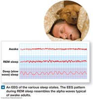
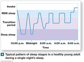
Arousal and Reticular Activating System (RAS)
Arousal is awakening from sleep, determined by interactions between reticular formation and cerebral cortex.
RAS projects to thalamic nuclei influencing large areas of cortex; stimulation produces widespread activation.
Sleep ends by any stimulus activating RAS; arousal occurs rapidly.
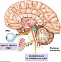
Regulation of Sleep–Wake Cycles
"Dueling" nuclei in brainstem: one stimulates RAS with NE (awake), another depresses RAS with serotonin (deep sleep).
Brain Chemistry and Behavior
Huntington’s disease: Destruction of ACh- and GABA-secreting neurons; motor and intellectual decline.
Serotonin: Affects sensory interpretation and emotional states; SSRIs prolong serotonin action.
Norepinephrine: Stimulation causes exhilaration; inhibition causes depression.
Dopamine: Controls intentional movements; deficiency causes Parkinson’s, excess may cause schizophrenia.
Effects of Aging on the Nervous System
Anatomical and Physiological Changes
Changes begin by age 30; 85% of people over 65 have changes in mental performance and CNS function.
Reduction in brain size and weight, decrease in cerebral cortex volume, narrower gyri, wider sulci, larger subarachnoid space.
Reduction in number of neurons; brain shrinkage linked to loss of cortical neurons.
Decrease in blood flow due to arteriosclerosis; increased risk of stroke.
Loss of synaptic connections; decline in neurotransmitter production.
Intracellular changes: lipofuscin, neurofibrillary tangles; extracellular changes: plaques (amyloid protein).
Functional Changes
Memory consolidation becomes more difficult; secondary memories harder to access.
Sensory acuity declines; reaction times slow; reflexes weaken; motor control precision decreases.
Most elderly retain functional ability; some develop senile dementia (memory loss, emotional disturbances, Alzheimer’s disease).
 Additional info: These notes are based on Chapter 16 of a college-level Anatomy & Physiology textbook, covering the autonomic nervous system, neurotransmitters, higher-order functions, and effects of aging. All images included are directly relevant to the adjacent content and reinforce key concepts.
