 Back
BackChapter 16: The Endocrine System – Structured Study Notes
Study Guide - Smart Notes
 Tailored notes based on your materials, expanded with key definitions, examples, and context.
Chapter 16: The Endocrine System
16.1 The Endocrine System as a Major Control System
The endocrine system is one of the body's two major control systems, working alongside the nervous system to coordinate and integrate the activity of most body cells. It uses hormones—chemical messengers transported in the blood—to influence metabolic activities. Endocrine responses are slower but longer-lasting than nervous system responses.
Endocrinology: The study of hormones and endocrine organs.
Main functions: Reproduction, growth and development, maintenance of electrolyte, water, and nutrient balance, regulation of cellular metabolism and energy balance, mobilization of body defenses.
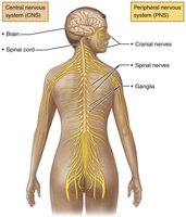
Comparison of Nervous and Endocrine Systems
The nervous system uses neurons and neurotransmitters for fast, short-lived responses, while the endocrine system uses glandular epithelia and hormones for slower, longer-lasting effects.
Nervous system: Acts at discrete locations, fast-acting, signal strength coded by action potential frequency.
Endocrine system: Acts at diffuse locations, long-lasting, signal strength coded by hormone concentration.
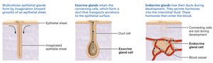
Endocrine vs. Exocrine Glands
Glands are classified based on their mode of secretion:
Exocrine glands: Produce nonhormonal substances (e.g., sweat, saliva), have ducts to carry secretion to membrane surfaces.
Endocrine glands: Produce hormones, ductless, secrete directly into extracellular fluid. Major glands include pituitary, thyroid, parathyroid, adrenal, and pineal glands.
Types of Chemical Messengers
Hormones: Long-distance chemical signals traveling in blood to target cells.
Autocrines: Affect the same cells that secrete them (local).
Paracrines: Affect neighboring cells (local).
Autocrines and paracrines are not considered part of the endocrine system.
16.2 Chemical Structure of Hormones
The chemical structure of a hormone determines its mechanism of action. There are two main classes:
Amino acid–based hormones: Includes derivatives, peptides, and proteins. Most are water-soluble (hydrophilic) and cannot cross the plasma membrane.
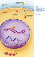
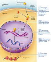
Steroid hormones: Synthesized from cholesterol, lipid-soluble (hydrophobic), can cross the plasma membrane. Includes gonadal and adrenocortical hormones.
16.3 Hormone Action and Target Cells
Hormones affect only cells with specific receptors—these are called target cells. Hormones alter target cell activity by increasing or decreasing the rates of normal cellular processes.
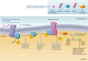
Hydrophilic (amino acid–based) hormones: Act via membrane receptors and second messenger systems.
Hydrophobic (steroid and thyroid) hormones: Act via intracellular receptors and direct gene activation.
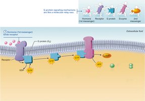
Second Messenger Systems
G protein-coupled receptors: Hormone binds to receptor, activates G protein, which then activates an enzyme to produce a second messenger (e.g., cAMP).
Cyclic AMP (cAMP) pathway: cAMP activates protein kinases, leading to cellular responses. Amplification occurs: one hormone can activate millions of protein kinases.
PIP2-calcium pathway: Hormone activates phospholipase C, splitting PIP2 into DAG and IP3, which activate kinases and release calcium from intracellular stores.

Direct Gene Activation
Lipid-soluble hormones: Bind to intracellular receptors, enter the nucleus, and activate specific genes to produce mRNA and proteins.
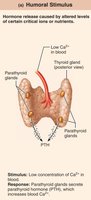
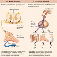
16.4 Stimuli for Hormone Release
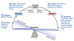
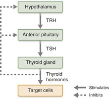
Hormone secretion is regulated by negative feedback mechanisms and is triggered by three types of stimuli:
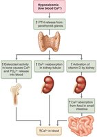
Humoral stimuli: Changing blood levels of ions/nutrients (e.g., Ca2+ stimulates parathyroid hormone release).
Neural stimuli: Nerve fibers stimulate hormone release (e.g., sympathetic stimulation of adrenal medulla).
Hormonal stimuli: Hormones stimulate other endocrine organs to release their hormones (e.g., hypothalamic-pituitary-target organ axis).

16.5 Hormone Receptors and Target Cell Activation
Cells respond to a hormone only if they have a receptor for it. The degree of target cell activation depends on:
Blood levels of the hormone
Number of receptors on/in the target cell
Affinity (strength) of binding between hormone and receptor
Up-regulation: Target cells increase receptor number in response to low hormone levels. Down-regulation: Target cells decrease receptor number in response to high hormone levels.
Half-Life, Onset, and Duration of Hormone Activity
Half-life: Time required for hormone level in blood to decrease by half.
Hormones are removed by liver, kidneys, or degraded by enzymes.
Onset and duration of hormone action vary by hormone type.
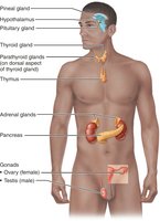
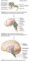
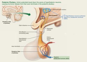
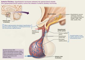
16.6 Hypothalamus and Pituitary Gland
The hypothalamus controls hormone release from the pituitary gland in two ways:
Posterior pituitary (neurohypophysis): Stores and releases neurohormones (oxytocin and ADH) produced by hypothalamic neurons.
Anterior pituitary (adenohypophysis): Glandular tissue that produces and releases six hormones regulated by hypothalamic releasing/inhibiting hormones via the hypophyseal portal system.
16.7 Thyroid Gland
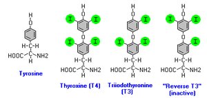
The thyroid gland is a butterfly-shaped organ located on the anterior trachea. It consists of follicular cells (produce thyroid hormone) and parafollicular cells (produce calcitonin).
Thyroid hormone (TH): Major metabolic hormone, produced as thyroxine (T4) and triiodothyronine (T3). Lipid-soluble, acts via intracellular receptors.
Effects: Increases basal metabolic rate, regulates growth and development, maintains blood pressure.
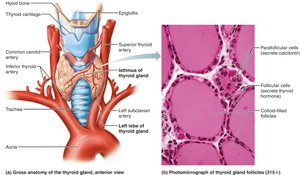
Calcitonin
Produced by parafollicular cells in response to high blood calcium.
Antagonist to parathyroid hormone (PTH); inhibits osteoclast activity and promotes calcium uptake into bone.
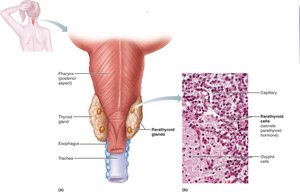
16.8 Parathyroid Glands
Parathyroid glands are small glands located on the posterior aspect of the thyroid gland. They secrete parathyroid hormone (PTH), the primary regulator of blood calcium levels.
PTH effects: Stimulates osteoclasts, enhances calcium reabsorption in kidneys, promotes activation of vitamin D for increased intestinal absorption of calcium.
16.9 Adrenal Glands
The adrenal glands are located atop the kidneys and consist of two regions:
Adrenal cortex: Produces corticosteroids (mineralocorticoids, glucocorticoids, gonadocorticoids).
Adrenal medulla: Produces catecholamines (epinephrine and norepinephrine) for the fight-or-flight response.
Adrenal Cortex Hormones
Mineralocorticoids (e.g., aldosterone): Regulate electrolyte balance, especially Na+ and K+.
Glucocorticoids (e.g., cortisol): Influence metabolism, help resist stress, maintain blood glucose.
Gonadocorticoids (androgens): Contribute to secondary sex characteristics and sex drive.
Adrenal Medulla Hormones
Catecholamines: Epinephrine and norepinephrine increase heart rate, blood pressure, and blood glucose during stress.
16.10 Pineal Gland
The pineal gland is located in the epithalamus and secretes melatonin, which regulates sleep-wake cycles and may have antioxidant and antigonadotropic effects.
16.11 Hormones Secreted by Other Organs
Many organs have endocrine functions in addition to their primary roles:
Pancreas: Produces insulin (lowers blood glucose) and glucagon (raises blood glucose).
Gonads: Ovaries produce estrogens and progesterone; testes produce testosterone.
Other organs: Heart (ANP, BNP), kidneys (erythropoietin, renin), skin (cholecalciferol), thymus (thymosins), adipose tissue (leptin, resistin, adiponectin), gastrointestinal tract (gastrin, ghrelin, secretin, CCK).
Summary Table: Major Endocrine Glands and Their Hormones
Gland | Main Hormones | Primary Functions |
|---|---|---|
Pituitary (anterior) | GH, TSH, ACTH, FSH, LH, PRL | Growth, metabolism, reproduction |
Pituitary (posterior) | Oxytocin, ADH | Uterine contraction, water balance |
Thyroid | T3, T4, Calcitonin | Metabolism, calcium regulation |
Parathyroid | PTH | Calcium homeostasis |
Adrenal cortex | Aldosterone, Cortisol, Androgens | Electrolyte balance, stress response, sex characteristics |
Adrenal medulla | Epinephrine, Norepinephrine | Fight-or-flight response |
Pineal | Melatonin | Sleep-wake cycles |
Pancreas | Insulin, Glucagon | Blood glucose regulation |
Ovaries | Estrogen, Progesterone | Female reproductive function |
Testes | Testosterone | Male reproductive function |
Additional info: These notes are based on the Marieb Human Anatomy & Physiology textbook, Twelfth Edition, and cover all major topics from Chapter 16: The Endocrine System, including hormone mechanisms, gland anatomy, and physiological effects. Images included are directly relevant to the explanation of each topic and reinforce key concepts for ANP college students.
