 Back
BackChapter 16: The Endocrine System – Structured Study Notes
Study Guide - Smart Notes
 Tailored notes based on your materials, expanded with key definitions, examples, and context.
Chapter 16: The Endocrine System
16.1 Overview of the Endocrine System
The endocrine system is one of the body's two major control systems, working alongside the nervous system to coordinate and integrate the activity of most body cells. It uses hormones—chemical messengers transported in the blood—to influence metabolic activities. Endocrine responses are generally slower but longer-lasting than those of the nervous system.
Endocrinology: The study of hormones and endocrine organs.
Major functions: Reproduction, growth and development, maintenance of electrolyte, water, and nutrient balance, regulation of cellular metabolism, and mobilization of body defenses.
Comparison of Nervous and Endocrine Systems
Nervous system: Uses electrical impulses and neurotransmitters, acts rapidly, effects are short-lived, and targets specific locations.
Endocrine system: Uses hormones, acts more slowly, effects are longer-lasting, and targets diffuse locations via the bloodstream.
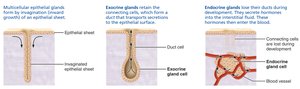
Types of Glands
Exocrine glands: Produce nonhormonal substances (e.g., sweat, saliva) and have ducts to carry secretions to membrane surfaces.
Endocrine glands: Ductless; secrete hormones directly into the extracellular fluid. Major glands include the pituitary, thyroid, parathyroid, adrenal, and pineal glands.

Types of Chemical Messengers
Hormones: Long-distance chemical signals that travel in blood to target cells.
Autocrines: Chemicals that exert effects on the same cells that secrete them.
Paracrines: Locally acting chemicals that affect neighboring cells.
Note: Autocrines and paracrines are local messengers and not considered part of the endocrine system.
16.2 Chemical Structure of Hormones
The chemical structure of a hormone determines how it acts. There are two main classes:
Amino acid–based hormones: Includes amino acid derivatives, peptides, and proteins. Most are water-soluble (hydrophilic) and cannot cross the plasma membrane (except thyroxine).
Steroid hormones: Synthesized from cholesterol, lipid-soluble (hydrophobic), and can cross the plasma membrane. Includes gonadal and adrenocortical hormones.
16.3 Mechanisms of Hormone Action
Hormones affect only target cells that have specific receptors. They alter target cell activity by increasing or decreasing the rates of normal cellular processes.
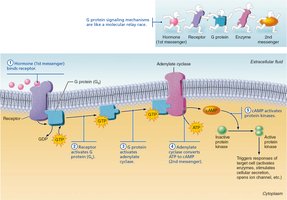
Water-soluble hormones (amino acid–based): Act on plasma membrane receptors, usually via second-messenger systems (e.g., cAMP, PIP2-calcium).
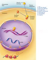
Lipid-soluble hormones (steroids and thyroid hormone): Act on intracellular receptors that directly activate genes.
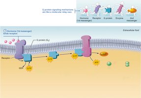
Second Messenger Systems
cAMP pathway: Hormone binds to receptor → activates G protein → activates adenylate cyclase → converts ATP to cAMP → activates protein kinases.
PIP2-calcium pathway: Hormone activates phospholipase C → splits PIP2 into DAG and IP3 → DAG activates protein kinases, IP3 releases Ca2+ from intracellular stores.
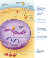
Direct Gene Activation
Lipid-soluble hormones diffuse into the cell, bind to intracellular receptors, and the hormone-receptor complex binds to DNA, initiating transcription and protein synthesis.
16.4 Control of Hormone Release
Hormone secretion is regulated by negative feedback mechanisms and is triggered by three types of stimuli:
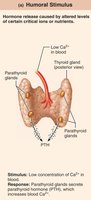
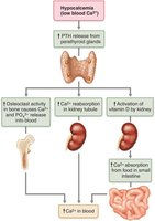
Humoral stimuli: Changes in blood levels of ions/nutrients (e.g., low Ca2+ stimulates PTH release).
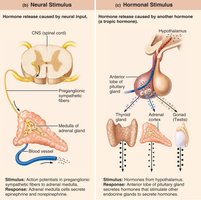
Neural stimuli: Nerve fibers stimulate hormone release (e.g., sympathetic stimulation of adrenal medulla).
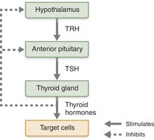
Hormonal stimuli: Hormones stimulate other endocrine organs to release their hormones (e.g., hypothalamic hormones regulate anterior pituitary).

16.5 Hormone Receptors and Target Cell Activation
Target cells must have specific receptors for a hormone to respond. The degree of activation depends on:
Blood levels of the hormone
Number of receptors on/in the target cell
Affinity (strength) of the binding between hormone and receptor
Up-regulation: Target cells form more receptors in response to low hormone levels. Down-regulation: Target cells lose receptors in response to high hormone levels.
Hormone Activity: Half-Life, Onset, and Duration
Half-life: Time required for hormone blood level to decrease by half.
Hormones are removed by the liver, kidneys, or degraded by enzymes.
Onset and duration of hormone action vary by hormone type.
16.6 The Hypothalamus and Pituitary Gland
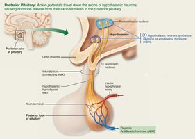
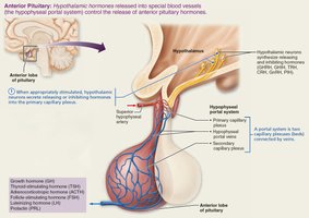
The hypothalamus controls the release of hormones from the pituitary gland in two ways. The pituitary has two major lobes:
Posterior pituitary (neurohypophysis): Composed of neural tissue; stores and releases oxytocin and ADH produced by the hypothalamus.
Anterior pituitary (adenohypophysis): Glandular tissue; produces and releases six hormones regulated by hypothalamic releasing and inhibiting hormones.
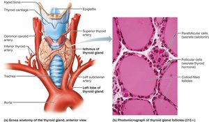
16.7 The Thyroid Gland
The thyroid gland is a butterfly-shaped organ located on the anterior trachea. It consists of two lobes connected by an isthmus and contains follicular cells (produce thyroid hormone) and parafollicular cells (produce calcitonin).
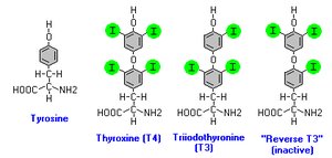
Thyroid hormone (TH): The body's major metabolic hormone, produced as T3 (triiodothyronine) and T4 (thyroxine). Increases basal metabolic rate, regulates tissue growth, and is critical for nervous system development.
Calcitonin: Produced by parafollicular cells; lowers blood calcium levels by inhibiting osteoclast activity.
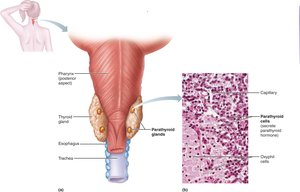
16.8 The Parathyroid Glands
Usually four small glands located on the posterior thyroid, the parathyroid glands secrete parathyroid hormone (PTH), the most important hormone in calcium homeostasis.
PTH: Increases blood calcium by stimulating osteoclasts, enhancing reabsorption in kidneys, and activating vitamin D for increased intestinal absorption.
16.9 The Adrenal Glands
The adrenal glands sit atop the kidneys and consist of two regions:
Adrenal cortex: Produces corticosteroids (mineralocorticoids, glucocorticoids, gonadocorticoids).
Adrenal medulla: Produces catecholamines (epinephrine and norepinephrine) for the fight-or-flight response.
Adrenal Cortex Hormones
Mineralocorticoids (e.g., aldosterone): Regulate Na+ and K+ balance.
Glucocorticoids (e.g., cortisol): Influence metabolism and help resist stress.
Gonadocorticoids: Weak androgens contributing to secondary sex characteristics.
Adrenal Medulla Hormones
Catecholamines: Epinephrine and norepinephrine increase heart rate, blood pressure, and blood glucose during stress.
16.10 The Pineal Gland
The pineal gland, located in the epithalamus, secretes melatonin, which regulates sleep-wake cycles and may have antioxidant and antigonadotropic effects.
16.11 Hormones Secreted by Other Organs
Several organs with other primary functions also secrete hormones:
Pancreas: Produces insulin (lowers blood glucose) and glucagon (raises blood glucose).
Gonads: Ovaries produce estrogens and progesterone; testes produce testosterone.
Other organs: Heart (ANP, BNP), kidneys (erythropoietin, renin), skin (cholecalciferol), thymus (thymosins), adipose tissue (leptin, resistin, adiponectin), GI tract (gastrin, secretin, CCK).
Pancreatic Hormones and Blood Glucose Regulation
Insulin: Promotes glucose uptake and storage, lowers blood glucose.
Glucagon: Stimulates glycogen breakdown and glucose release, raises blood glucose.
Summary Table: Major Endocrine Glands and Their Hormones
Gland | Hormone(s) | Main Function(s) |
|---|---|---|
Pituitary (anterior) | GH, TSH, ACTH, FSH, LH, PRL | Growth, metabolism, stress, reproduction |
Pituitary (posterior) | Oxytocin, ADH | Uterine contraction, water balance |
Thyroid | T3, T4, Calcitonin | Metabolism, calcium regulation |
Parathyroid | PTH | Calcium homeostasis |
Adrenal cortex | Aldosterone, Cortisol, Androgens | Electrolyte balance, stress response, sex characteristics |
Adrenal medulla | Epinephrine, Norepinephrine | Fight-or-flight response |
Pineal | Melatonin | Sleep-wake cycles |
Pancreas | Insulin, Glucagon | Blood glucose regulation |
Gonads | Estrogens, Progesterone, Testosterone | Reproduction, secondary sex characteristics |
