 Back
Chapter 17: Blood – Structure, Function, and Disorders
Study Guide - Smart Notes
 Tailored notes based on your materials, expanded with key definitions, examples, and context.
Blood Composition
Main Components of Blood
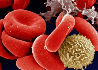
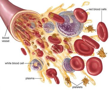
Blood is a specialized fluid connective tissue composed of plasma and formed elements. The formed elements include erythrocytes (red blood cells), leukocytes (white blood cells), and platelets.
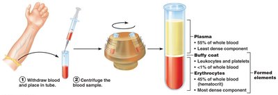
Plasma: The liquid matrix, making up about 55% of blood volume, contains water, proteins, nutrients, electrolytes, and waste products.
Erythrocytes (RBCs): Responsible for oxygen transport.
Leukocytes (WBCs): Provide immune defense.
Platelets: Essential for blood clotting (coagulation).
Hematocrit and Blood Separation
Hematocrit is the percentage of blood volume occupied by erythrocytes. Typical values are 47% ± 5% for males and 42% ± 5% for females. Leukocytes and platelets form a thin 'buffy coat' (~1%), and plasma makes up the remainder.
Physical Characteristics and Functions of Blood
Physical Properties
Blood is a sticky, opaque fluid with a pH of 7.35–7.45 and a temperature of about 38°C. It constitutes approximately 8% of body weight, with average volumes of 5–6 L for males and 4–5 L for females.
Functions of Blood
Blood performs three primary functions: distribution, regulation, and protection.
Distribution: Transports oxygen, nutrients, metabolic wastes, and hormones.
Regulation: Maintains body temperature, pH balance, and fluid volume.
Protection: Prevents blood loss via clotting and defends against infection through immune cells and proteins.
Blood Plasma
Composition of Plasma
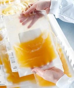
Plasma is 90% water and contains over 100 dissolved solutes, including plasma proteins (albumin, globulins, fibrinogen), nutrients, electrolytes, respiratory gases, and hormones.
Formed Elements
Types and Characteristics
Formed elements include erythrocytes, leukocytes, and platelets. Only leukocytes are complete cells; erythrocytes lack nuclei and organelles, and platelets are cell fragments. Most formed elements originate in bone marrow and have limited lifespans.
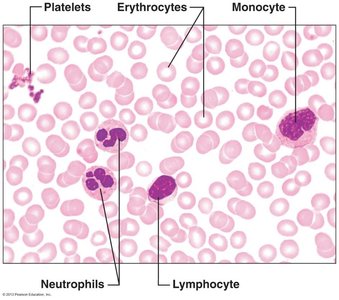
Erythrocytes (Red Blood Cells)
Structure and Function
Erythrocytes are biconcave, anucleate cells filled with hemoglobin for gas transport. Their shape increases surface area for efficient gas exchange and flexibility for passage through capillaries.
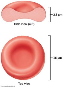
Hemoglobin Structure and Function
Hemoglobin is a protein composed of two alpha and two beta globin chains, each with a heme group containing iron. Each hemoglobin molecule can bind four oxygen molecules.
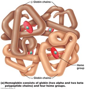
Oxygen loading: Forms oxyhemoglobin (ruby red).
Oxygen unloading: Forms deoxyhemoglobin (dark red).
CO2 loading: Forms carbaminohemoglobin (carries 20% of CO2).
Hematopoiesis
Blood Cell Formation
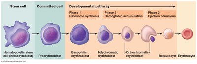
Hematopoiesis is the process of blood cell formation, occurring in red bone marrow. Hemocytoblasts (hematopoietic stem cells) differentiate into various blood cells under hormonal and growth factor control.
Erythropoiesis
Erythropoiesis is the production of red blood cells, involving transformation from hemocytoblast to proerythroblast, then through several stages to mature erythrocyte. The process takes about 15 days.
Phase 1: Ribosome synthesis
Phase 2: Hemoglobin accumulation
Phase 3: Ejection of nucleus and formation of reticulocyte
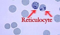
Regulation of Erythropoiesis
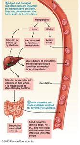
Regulation depends on hormonal control (primarily erythropoietin, EPO, released by kidneys in response to hypoxia) and adequate nutrients (amino acids, iron, vitamin B12, folic acid).
Fate and Destruction of Erythrocytes
Lifecycle and Breakdown
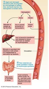
RBCs live 100–120 days. Old cells are engulfed by macrophages in the spleen, liver, and bone marrow. Hemoglobin is broken down: iron is salvaged, heme is degraded to bilirubin, and globin is metabolized to amino acids.
Erythrocyte Disorders
Anemia
Anemia is a condition of abnormally low oxygen-carrying capacity, resulting in fatigue, paleness, shortness of breath, and chills. Causes include insufficient erythrocytes, low hemoglobin content, and abnormal hemoglobin.

Hemorrhagic anemia: Blood loss.
Hemolytic anemia: RBCs rupture prematurely.
Aplastic anemia: Bone marrow destruction.
Iron-deficiency anemia: Inadequate iron intake or absorption.
Pernicious anemia: Vitamin B12 deficiency.
Thalassemias: Faulty globin chain.
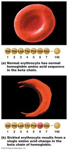
Sickle-cell anemia: Abnormal hemoglobin causes sickle-shaped RBCs.
Polycythemia
Polycythemia is an excess of RBCs, increasing blood viscosity. Causes include bone marrow cancer, high altitude, increased EPO production, and blood doping.
Leukocytes (White Blood Cells)
Types and Functions
Leukocytes make up less than 1% of blood volume and are crucial for immune defense. They can leave capillaries (diapedesis) and move toward damaged cells (chemotaxis).
Granulocytes
Granulocytes include neutrophils, eosinophils, and basophils. They have lobed nuclei and cytoplasmic granules.
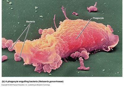
Neutrophils: Most numerous, phagocytic, "bacteria slayers."
Eosinophils: Digest parasitic worms, modulate immune response.
Basophils: Release histamine, similar to mast cells.
Agranulocytes
Agranulocytes include lymphocytes and monocytes, which lack visible granules.
Lymphocytes: Crucial to immunity; T cells attack infected cells, B cells produce antibodies.
Monocytes: Largest WBCs; differentiate into macrophages for phagocytosis and immune activation.
Leukopoiesis and Leukocyte Disorders
Leukopoiesis
Leukopoiesis is the production of WBCs, stimulated by interleukins and colony-stimulating factors. All leukocytes originate from hemocytoblasts.
Leukocyte Disorders
Leukopenia: Abnormally low WBC count.
Leukemia: Cancerous proliferation of WBCs.
Infectious mononucleosis: Excessive agranulocytes due to Epstein-Barr virus.
AIDS: HIV attacks helper T-cells, crippling immune response.
Platelets and Hemostasis
Platelet Structure and Function
Platelets are fragments of megakaryocytes, regulated by thrombopoietin. They form temporary plugs to seal vessel breaks and contain chemicals for clotting.
Hemostasis
Hemostasis is the process of stopping bleeding, involving three steps:
Vascular spasm: Vasoconstriction reduces blood loss.
Platelet plug formation: Platelets adhere to exposed collagen, release chemicals to recruit more platelets.
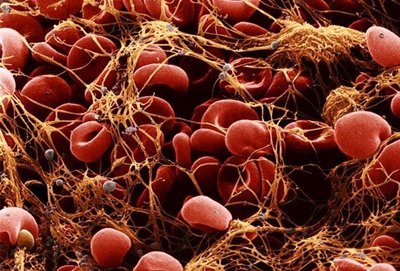
Coagulation: Clotting factors transform blood from liquid to gel, reinforcing the plug with fibrin threads.
Coagulation Cascade
Coagulation involves three phases:
Formation of prothrombin activator (intrinsic and extrinsic pathways).
Conversion of prothrombin to thrombin.
Thrombin catalyzes the joining of fibrinogen to form a fibrin mesh.
Phase | Key Event |
|---|---|
1 | Prothrombin activator formed |
2 | Prothrombin converted to thrombin |
3 | Fibrinogen converted to fibrin mesh |
Clot Retraction and Repair
Platelets contract to compact the clot and bring vessel edges together. Growth factors stimulate repair of vessel walls and endothelial lining.
Fibrinolysis
Fibrinolysis removes clots after healing. Plasminogen is converted to plasmin, which digests fibrin.
Blood Groups and Transfusions
Blood Group Antigens
RBC membranes bear glycoprotein antigens (agglutinogens) used to classify blood into groups. The ABO and Rh groups are most clinically significant.
ABO: Types A, B, AB, O based on presence/absence of A and B antigens.
Rh: Rh+ indicates presence of D antigen; anti-Rh antibodies form after exposure.
Transfusion Reactions
Transfusion of incompatible blood causes agglutination and hemolysis, leading to diminished oxygen-carrying capacity and possible renal failure.
Restoring Blood Volume
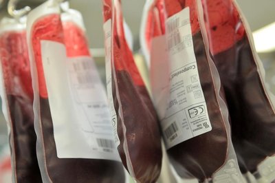
Blood volume can be restored with saline, electrolyte solutions, or plasma expanders to prevent shock from blood loss.
Summary Table: Blood Components and Functions
Component | Main Function | Key Features |
|---|---|---|
Plasma | Transport, regulation | 90% water, proteins, nutrients, electrolytes |
Erythrocytes | Oxygen transport | Biconcave, hemoglobin-rich, anucleate |
Leukocytes | Immune defense | Granulocytes & agranulocytes, complete cells |
Platelets | Clotting | Cell fragments, contain clotting chemicals |
Key Equations
Hematocrit:
Oxygen Transport:
Coagulation:
Additional info: Academic context was added to clarify the structure and function of blood components, the process of hematopoiesis, and the clinical significance of blood disorders and transfusions.
