 Back
BackChapter 17: The Special Senses – Olfaction, Gustation, Hearing, Equilibrium, and Vision
Study Guide - Smart Notes
 Tailored notes based on your materials, expanded with key definitions, examples, and context.
The Special Senses
Overview of Sensory Types
The human body possesses two main types of senses: general senses and special senses. General senses include pain, temperature, touch, pressure, vibration, and proprioception. Special senses are more complex and include olfaction (smell), vision, gustation (taste), equilibrium (balance), and hearing. Specialized sensory receptors provide the central nervous system (CNS) with information about the external and internal environment.
Olfaction (Smell)
Anatomy of the Olfactory Organ
Olfaction is the sense of smell, occurring in the nasal cavity. The olfactory organ consists of two major parts: the olfactory epithelium and the lamina propria. The olfactory epithelium covers the inferior surface of the cribriform plate, the superior portion of the perpendicular plate, and the superior nasal conchae of the ethmoid bone. It contains olfactory receptor cells, supporting cells, and basal cells. The lamina propria is an underlying layer of areolar connective tissue containing olfactory glands that produce mucus.
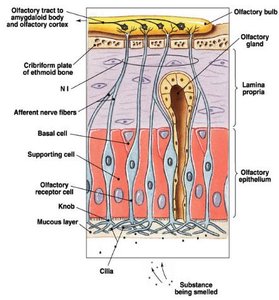
Olfactory Receptors and Signal Transduction
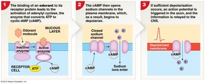
Olfactory receptors are highly modified neurons whose dendritic bulbs extend into the surrounding mucus. Odorant molecules dissolve in the mucus and bind to odorant-binding proteins (G proteins) on the dendrites, activating a second messenger system. Adenylyl cyclase converts ATP to cAMP, which opens sodium channels, causing depolarization and triggering an action potential. This information is relayed to the CNS.
Odorant molecules: Dissolved organic molecules that stimulate olfactory receptors.
Second Messenger System: Involves G proteins, adenylyl cyclase, cAMP, and sodium channels.
Olfactory Discrimination
Humans can distinguish between 2,000–4,000 odorants. There are populations of olfactory neurons with distinct sensitivities, and more than 50 primary smells are interpreted based on the overall pattern of receptor activity. Olfactory neurons are replaced frequently, but their total number declines with age.
Gustation (Taste)
Anatomy of Taste Buds
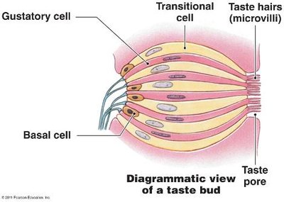
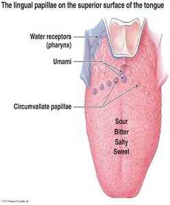
Gustation is the sense of taste, mediated by gustatory receptors found in taste buds. Taste buds are distributed over the superior surface of the tongue and adjacent portions of the pharynx and larynx. Each taste bud contains about 40 slender, spindle-shaped cells, including gustatory receptors, basal cells (stem cells), and transitional cells. Microvilli (taste hairs) extend into the surrounding fluid through a taste pore.
Lingual Papillae
The tongue contains four types of epithelial projections called lingual papillae:
Filiform papillae: Provide friction, help move objects in the mouth, no taste buds.
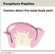
Fungiform papillae: Each contains about 5 taste buds, scattered on the tongue, higher concentrations on sides and tip.
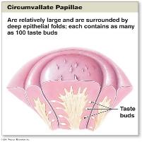
Vallate (circumvallate) papillae: Each contains about 100 taste buds, form a "V" near the posterior margin.
Foliate papillae: Found in folds along the lateral margin of the tongue, contain taste buds.
Other Taste Sensations
In addition to the primary taste sensations (sweet, salty, sour, bitter), humans can detect umami (a pleasant, savory taste imparted by glutamate, found in foods like beef broth and parmesan cheese) and water receptors (affecting water balance and blood volume, mostly on the pharynx).
Gustatory Discrimination and Pathway
Primary taste sensations are detected by different mechanisms:
Sweet: G protein–coupled receptors
Salty: Sodium ions diffuse into receptor cell via sodium leak channels
Sour: Hydrogen ions diffuse into receptor cell via sodium leak channels
Bitter: G protein–coupled receptors
Gustatory receptors depolarize and synapse with sensory neurons forming the facial (VII), glossopharyngeal (IX), and vagus (X) nerves. These nerves synapse in the medulla oblongata, then the thalamus, and finally the gustatory cortex of the brain.
The Ear: Hearing and Equilibrium
Anatomy of the Ear
The ear is divided into three regions: external ear, middle ear, and inner ear. The external ear collects and directs sound waves to the eardrum. The middle ear amplifies sound waves and transmits them to the inner ear. The inner ear contains the bony and membranous labyrinths, which house receptors for equilibrium and hearing.
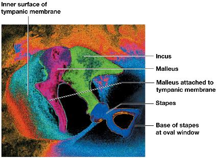
Middle Ear Structures
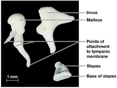
The middle ear contains the tympanic cavity and three auditory ossicles (malleus, incus, stapes) that transmit and amplify sound vibrations. The pharyngotympanic tube connects the middle ear with the nasopharynx, permitting equalization of pressure.
Inner Ear Structures
The inner ear consists of the bony labyrinth (protects membranes) and membranous labyrinth (fluid-filled tubes). It contains perilymph and endolymph, and houses the semicircular canals (balance), vestibule (linear acceleration and gravity), and cochlea (hearing).
Equilibrium
Equilibrium sensations are detected by hair cells in the vestibular complex (vestibule and semicircular canals). Hair cells in semicircular ducts detect rotational movement, while those in the vestibule detect body position with respect to gravity and acceleration.
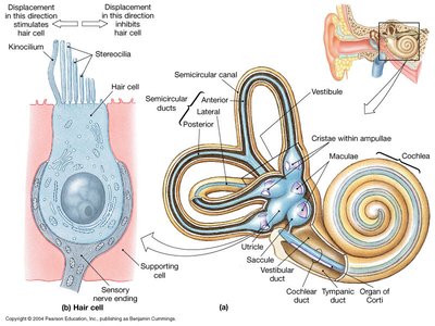
Vestibule and Macula
The macula is an oval structure in the saccule and utricle containing hair cells. The otolithic membrane is a gelatinous structure with otoliths (calcium carbonate crystals) that move with gravity, bending the stereocilia of hair cells and releasing neurotransmitter onto sensory nerve endings.

Hearing: Cochlea and Spiral Organ
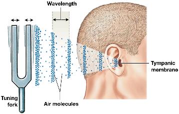
Sound waves are converted into mechanical movements by vibration of the tympanic membrane. Auditory ossicles conduct vibrations to the internal ear, where they are converted to pressure waves in fluid and detected by hair cells in the cochlear duct. The cochlear duct is filled with endolymph and lies between two chambers filled with perilymph (vestibular and tympanic ducts).
Hearing Process
Sound waves arrive at the tympanic membrane and make it vibrate.
Movement of the tympanic membrane displaces the auditory ossicles, amplifying the vibration.
Movement of the stapes at the oval window produces pressure waves in the perilymph of the vestibular duct.
Pressure waves distort the basilar membrane on their way to the round window of the tympanic duct.
Vibration of the basilar membrane causes hair cells to vibrate against the tectorial membrane.
Information about stimulation is relayed to the CNS over the cochlear nerve, a division of the vestibulocochlear nerve.
The Eye: Vision
Structure of the Eye
The eye is a hollow spheroid filled with fluid, surrounded by three layers: fibrous tunic, vascular tunic, and retina (neural tunic). The anterior cavity contains aqueous humor, while the posterior cavity contains the vitreous body.
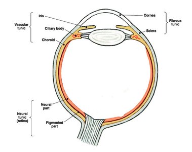
Fibrous Tunic
The fibrous tunic consists of the sclera (white of the eye) and cornea (transparent, continuous with sclera). It provides mechanical support, protection, and assists in focusing.
Vascular Tunic
The vascular tunic contains blood vessels, lymphatics, and intrinsic muscles. It regulates light entry, secretes and reabsorbs aqueous humor, and controls lens shape for focusing. The iris, pupil, ciliary body, and choroid are components of the vascular tunic.
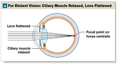
Lens and Refraction
The lens is a transparent, flexible disc that focuses visual images on photoreceptors by changing shape. Refraction occurs when light passes from one medium to another, bending toward the focal point on the retina. Focal distance is determined by object distance and lens shape.
Accommodation
Accommodation is the process of changing the shape of the lens to focus images on the retina. Ciliary muscle contraction makes the lens rounder for close vision, while relaxation flattens the lens for distant vision.
Retina (Neural Tunic)
The retina is the innermost layer, containing photoreceptors (rods and cones), supporting cells, and neurons. Rods enable vision in dim light, while cones provide color vision and are concentrated at the fovea of the macula lutea.
Visual Pathway
The visual pathway begins at photoreceptors, which synapse with bipolar cells, then ganglion cells. Axons of ganglion cells form the optic nerve, which relays information to the thalamus and occipital cortex.
Special Sense | Receptor Type | Main Structures | Pathway to CNS |
|---|---|---|---|
Olfaction | Olfactory receptor cells | Olfactory epithelium, lamina propria | Olfactory nerve → CNS |
Gustation | Gustatory receptor cells | Taste buds, lingual papillae | Facial, glossopharyngeal, vagus nerves → Medulla → Thalamus → Gustatory cortex |
Hearing | Hair cells | Cochlea, spiral organ | Cochlear nerve → Medulla → Inferior colliculus → Thalamus → Auditory cortex |
Equilibrium | Hair cells | Vestibule, semicircular canals | Vestibular nerve → CNS |
Vision | Rods and cones | Retina, lens, cornea | Optic nerve → Thalamus → Occipital cortex |
Additional info: Academic context was added to clarify the function and structure of each special sense, as well as the pathways by which sensory information is relayed to the CNS.
