 Back
BackChapter 18: The Cardiovascular System – The Heart
Study Guide - Smart Notes
 Tailored notes based on your materials, expanded with key definitions, examples, and context.
Heart Anatomy & Characteristics
Size, Location, and Structure
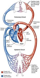
The heart is a muscular organ roughly the size of a closed fist (250–350 g), located in the thoracic cavity just left of the midline. It is divided into right and left sides, each serving distinct circulatory functions. - Right side: Pulmonary circuit – pumps blood to the lungs for oxygenation. - Left side: Systemic circuit – pumps oxygenated blood to body tissues. - Atria: Two superior chambers (right and left) act as receiving chambers for blood returning from the body and lungs. - Ventricles: Two inferior chambers (right and left) serve as pumping chambers, sending blood out to the lungs and body. - Pericardium: The heart is enclosed in a membrane-lined cavity within the thoracic cavity. - Heart wall layers:
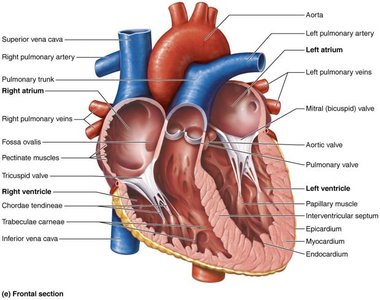
Epicardium: Thin outer membrane.
Myocardium: Thickest layer, composed of cardiac muscle.
Endocardium: Thin inner membrane lining chambers and covering valves; continuous with inner membrane of blood vessels.
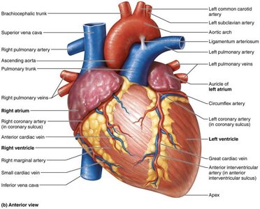
Chambers & Vessels
The heart is internally divided by septa: - Interatrial septum: Divides the atria. - Interventricular septum: Divides the ventricles. Auricles are extensions of the atria that increase their volume. Right atrium (RA): Receives blood from:
Superior vena cava (from body superior to diaphragm)
Inferior vena cava (from body inferior to diaphragm)
Coronary sinus (from myocardium)
Left atrium (LA): Receives blood from four pulmonary veins (from lungs). Ventricles: Have thicker walls than atria, as they discharge blood from the heart.
Right ventricle (RV): Pumps blood through pulmonary trunk into right and left pulmonary arteries, to the lungs.
Left ventricle (LV): Pumps blood through the aorta to the rest of the body.

Heart Valves
Atrioventricular (AV) and Semilunar Valves
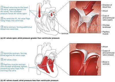
Heart valves ensure one-way flow of blood and prevent backflow. They open and close in response to pressure changes in the chambers. Atrioventricular (AV) Valves: Located between atria and ventricles.
Tricuspid valve: Three cusps; between RA & RV.
Bicuspid (mitral) valve: Two cusps; between LA & LV.
Chordae tendineae: CT cords anchor valves to ventricular walls, preventing hyperextension into the atria.
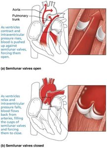
Semilunar Valves: Located between ventricles and arteries exiting the heart.
Aortic valve: Three cusps; between LV & aorta.
Pulmonary valve: Three cusps; between RV & pulmonary trunk.
Blood Flow Through the Heart
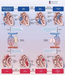
Pulmonary and Systemic Circulations
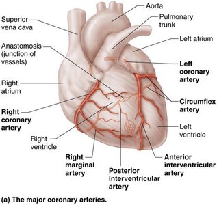
The heart supports two major circulatory pathways: pulmonary and systemic. Both circuits carry the same volume of blood but operate under different pressures. - RV → lungs: Low pressure. - LV → body: High pressure (LV wall is three times thicker than RV). Coronary circulation: Supplies oxygenated blood to the myocardium.
Left and right coronary arteries: Arise from the aorta and branch to supply the heart.
Anterior interventricular artery: Supplies blood to the interventricular septum and anterior walls of both ventricles.
Circumflex artery: Supplies blood to the LA and posterior walls of LV.
Cardiac Muscle and Conduction System
Cardiac Muscle Cell Types
Cardiac muscle contraction is initiated by action potentials. There are two types of cardiac muscle cells: - Contractile cells: Responsible for contractions; majority of cardiac muscle cells. - Pacemaker cells: Noncontractile; generate and send impulses to set the heart’s rhythm (autorhythmic). Note: The nervous system does not directly initiate these impulses, unlike skeletal muscle.
Intrinsic Conduction System
The heart’s rhythm is set by a specialized conduction system:
SA (sinoatrial) node: Pacemaker in wall of RA; generates 75 impulses/min (sinus rhythm).
AV (atrioventricular) node: Superior to tricuspid valve; impulses pause to allow atrial contraction.
AV bundle: In superior interventricular septum; electrical connection between atria and ventricles.
Subendocardial conducting network (Purkinje fibers): Completes pathway through apex and ventricle walls.
Electrocardiogram (ECG)
ECG Waves and Cardiac Cycle
An electrocardiogram (ECG) records the electrical impulses that cause contraction and relaxation of heart muscle. - P wave: Atrial contraction (depolarization). - QRS complex: Ventricular contraction (depolarization); obscures atrial relaxation. - T wave: Ventricular relaxation (repolarization). 
Cardiac Cycle
Systole, Diastole, and Blood Flow
The cardiac cycle includes all events associated with blood flow during one complete heartbeat. - Systole: Contraction phase. - Diastole: Relaxation phase. - Duration: Lasts about 0.8 seconds. - Pressure and volume changes: Blood flow is controlled by pressure changes; blood flows down pressure gradients through available openings.
Cardiac Output, Heart Rate, and Stroke Volume
Definitions and Formula
Cardiac output (CO): Volume of blood pumped out of each ventricle in one minute.
Stroke volume (SV): Volume of blood pumped out of each ventricle in one beat.
Heart rate (HR): Number of beats per minute.
Formula: Example:
Entire blood supply passes through each side of the heart once per minute.
CO varies directly with HR and SV.
Cardiac reserve is the difference between resting and maximum CO.
Regulation of Heart Rate
Autonomic Nervous System (ANS) Regulation
In healthy individuals, SV remains relatively constant. If SV decreases (e.g., due to blood loss or weakened heart muscle), HR and contractility increase to maintain CO. - Sympathetic nervous system: Releases norepinephrine, causing SA node to fire more rapidly and increasing HR during emotional or physical stress. - Parasympathetic nervous system: Slows HR when stressors subside.
Other Regulators of Heart Rate
Hormones: Epinephrine raises HR and contractility; thyroid hormone raises HR and metabolic rate, enhancing epinephrine effects.
Ion imbalances: Pose a threat to proper heart function.
Age: Resting HR is fastest in fetuses (140–160 bpm) and declines throughout life.
Gender: Resting HR is faster in females (72–80 bpm) than males (64–72 bpm).
Exercise: Raises HR via sympathetic nervous system intervention.
Summary Table: Heart Chambers, Valves, and Major Vessels
Chamber | Receives Blood From | Pumps Blood To | Associated Valve |
|---|---|---|---|
Right Atrium | Superior/Inferior Vena Cava, Coronary Sinus | Right Ventricle | Tricuspid Valve |
Right Ventricle | Right Atrium | Pulmonary Trunk | Pulmonary Valve |
Left Atrium | Pulmonary Veins | Left Ventricle | Bicuspid (Mitral) Valve |
Left Ventricle | Left Atrium | Aorta | Aortic Valve |
Summary Table: Major Coronary Arteries and Veins
Artery | Supplies |
|---|---|
Left Coronary Artery | LA, LV, Interventricular Septum |
Right Coronary Artery | RA, RV |
Circumflex Artery | LA, Posterior LV |
Anterior Interventricular Artery | Anterior Walls of Both Ventricles |
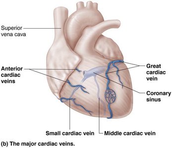
Vein | Drains |
|---|---|
Great Cardiac Vein | Anterior Heart |
Middle Cardiac Vein | Posterior Heart |
Small Cardiac Vein | Right Heart |
Coronary Sinus | Collects from all cardiac veins |
