 Back
BackChapter 18: The Heart – Structure, Function, and Circulation
Study Guide - Smart Notes
 Tailored notes based on your materials, expanded with key definitions, examples, and context.
The Heart as Part of the Cardiovascular System
Components and Functions
The heart is a central organ in the cardiovascular system, responsible for pumping blood throughout the body. The cardiovascular system consists of three main components:
Heart: Acts as a pump to circulate blood.
Blood vessels: Serve as delivery routes for blood.
Blood: Serves as the delivery medium for oxygen, nutrients, carbon dioxide, and wastes.
The heart ensures that oxygen and nutrients reach cells, while carbon dioxide and wastes are removed.
Location and Structure of the Heart
Position in the Mediastinum
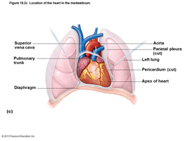
The heart is located in the mediastinum, a central compartment in the thoracic cavity. It lies between the lungs, behind the sternum, and above the diaphragm. The apex of the heart points downward and to the left, while the base anchors the heart and faces toward the right shoulder.
Size: About the size of a fist (~300 g).
Point of maximum impulse: Between the 5th and 6th ribs at the midclavicular line.
Surrounding Structures
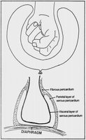
The heart is surrounded by the pericardium, a double-layered sac that encloses the heart and part of the great vessels.
Fibrous pericardium: Dense irregular connective tissue that protects, anchors, and prevents overstretching.
Serous pericardium: Simple squamous epithelium with two layers: parietal (lines inside fibrous pericardium) and visceral (lines heart, also called epicardium). Produces serous fluid to prevent friction.
Layers of the Heart Wall
Structural Layers
The heart wall consists of three distinct layers, each with specialized functions:
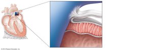
Epicardium: External layer, simple squamous epithelium.
Myocardium: Middle contractile layer, composed of cardiac muscle cells, making up 95% of the heart wall.
Endocardium: Inner layer, simple squamous epithelium, lines chambers and valves, continuous with endothelium of blood vessels, minimizes friction.
Heart Chambers and Basic Anatomy
Chamber Structure
The heart has four chambers:
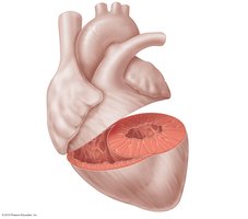
Atria: Superior chambers, divided by the interatrial septum. They are receiving chambers with lower pressure and thinner myocardium.
Ventricles: Inferior chambers, divided by the interventricular septum. They act as pumps with higher pressure and thicker myocardium.
Key anatomical features include:
Fossa ovalis: Remnant of the foramen ovale, a fetal shunt.
Papillary muscles and chordae tendineae: Attach to valves and prevent backflow during ventricular contraction.
Sulci: Grooves marking chamber separation (atrioventricular, anterior interventricular, posterior interventricular).
Circuits of the Heart
Pulmonary and Systemic Circuits
The heart functions as two pumps:
Pulmonary circuit: Right heart → lungs → left heart. Delivers O2-poor blood to lungs for gas exchange; returns O2-rich blood to left heart.
Systemic circuit: Left heart → body → right heart. Delivers O2-rich blood to body; returns O2-poor blood to right heart.
Both ventricles pump the same amount of blood, but the left ventricle has a thicker wall due to higher systemic pressure.
Fibrous Skeleton of the Heart
Structure and Function
The fibrous skeleton is dense connective tissue running through cardiac muscle. It supports heart valves and great vessels, serves as a point of attachment for cardiac muscle, and electrically insulates atria from ventricles, ensuring proper timing of contractions.
Pathway of Blood Through the Heart
Sequential Flow
Blood follows a specific pathway through the heart and body:
SVC and IVC → Right Atrium (RA)
Tricuspid valve → Right Ventricle (RV)
Pulmonic valve → Pulmonary Trunk
Left & Right Pulmonary arteries → Lungs (gas exchange)
Left & Right Pulmonary veins → Left Atrium (LA)
Mitral valve → Left Ventricle (LV)
Aortic valve → Aorta
Body capillaries (gas exchange)
Coronary Circulation
Blood Supply to the Heart
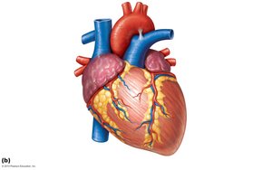
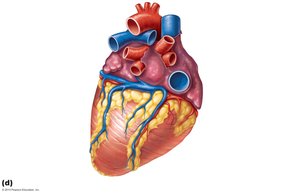
Coronary arteries branch from the aorta and supply blood to cardiac muscle tissue, which has high energy requirements and can only produce ATP aerobically. Blockage leads to myocardial infarction (heart attack), resulting in muscle death and replacement by scar tissue. Anastomoses provide multiple pathways for blood flow.
Heart Valves
Types and Functions
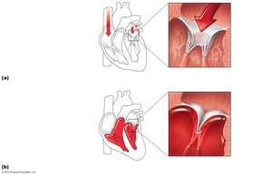
Heart valves ensure unidirectional blood flow by opening and closing in response to pressure changes:
Atrioventricular (AV) valves: Tricuspid (RA to RV), Mitral (LA to LV).
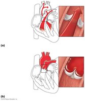
Semilunar valves: Pulmonic (RV to pulmonary trunk), Aortic (LV to aorta).
Blood flows from atria → ventricles → vessels.
Valve Functioning
Valve operation depends on pressure differences:
Atrial pressure > ventricular pressure: AV valves open.
Ventricular pressure > atrial pressure: AV valves close.
Ventricular pressure > aortic pressure: Aortic valve opens.
Aortic pressure > ventricular pressure: Aortic valve closes.
Summary Table: Heart Valves and Their Functions
Valve | Location | Function |
|---|---|---|
Tricuspid (AV) | RA to RV | Prevents backflow into RA |
Mitral (AV) | LA to LV | Prevents backflow into LA |
Pulmonic (Semilunar) | RV to Pulmonary Trunk | Prevents backflow into RV |
Aortic (Semilunar) | LV to Aorta | Prevents backflow into LV |
Key Terms and Concepts
Myocardial infarction: Death of cardiac muscle due to lack of oxygen.
Anastomosis: Connection between two or more arteries.
Chordae tendineae: Tendinous cords preventing valve prolapse.
Papillary muscles: Muscles anchoring chordae tendineae.
Relevant Equations
Cardiac Output
Cardiac output is the volume of blood pumped by the heart per minute:
Blood Pressure
Blood pressure is determined by cardiac output and resistance:
Conclusion
The heart is a complex organ with specialized structures and functions that ensure efficient circulation of blood throughout the body. Understanding its anatomy, circuits, and valve mechanisms is essential for comprehending cardiovascular physiology.
