 Back
BackChapter 19: Blood – Structure, Function, and Disorders
Study Guide - Smart Notes
 Tailored notes based on your materials, expanded with key definitions, examples, and context.
Blood: Functions and General Characteristics
Functions of Blood
Blood is a vital connective tissue that performs several essential functions in the human body, including transport, protection, and regulation.
Transport and Distribution: Blood carries oxygen (O2) from the lungs to tissues, carbon dioxide (CO2) from tissues to the lungs, nutrients from the digestive system, hormones from endocrine glands, and metabolic heat.
Protection: Platelets and clotting factors prevent blood loss (hemostasis), while leukocytes and antibodies fight infection.
Regulation: Blood maintains ion and fluid balance, buffers acids and bases to stabilize pH, distributes heat, and helps regulate blood pressure.
General Characteristics of Blood
Blood is more viscous and dense than water, with a pH of approximately 7.35–7.45 (slightly alkaline) and a temperature around 38°C (100°F). Blood volume depends on body mass, averaging 5–6 liters in males and 4–5 liters in females.
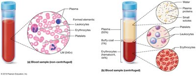
Components of Whole Blood
Whole blood consists of two main components: formed elements (cells and cell fragments) and plasma (the liquid portion).
Formed Elements: Red blood cells (RBCs), white blood cells (WBCs), and platelets.
Plasma: A yellowish extracellular fluid that suspends blood solutes and carries heat.
Blood Plasma: Composition and Functions
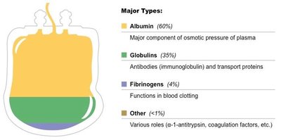
Major Components of Plasma
Blood plasma is composed of water, proteins, electrolytes, nutrients, wastes, and gases. Plasma proteins are primarily synthesized in the liver and remain in the circulatory system.
Albumin: Maintains osmotic pressure and acts as a buffer for blood pH.
Globulins: Include antibodies (immunoglobulins) and transport proteins.
Fibrinogen: Essential for blood clotting.
Other: Hormones, enzymes, and other proteins.
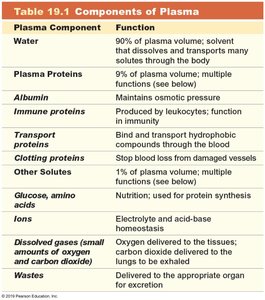
Plasma Component | Function |
|---|---|
Water | Solvent; dissolves and transports solutes |
Plasma Proteins | Multiple functions (osmotic pressure, immunity, transport, clotting) |
Albumin | Maintains osmotic pressure |
Immune proteins | Produced by leukocytes; immunity |
Transport proteins | Bind and transport hydrophobic compounds |
Clotting proteins | Stop blood loss from damaged vessels |
Glucose, amino acids, ions | Used for protein synthesis and acid-base homeostasis |
Dissolved gases | Oxygen and carbon dioxide transport |
Wastes | Transported for excretion |
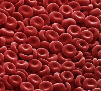
Formed Elements: Erythrocytes (Red Blood Cells)
Structure and Function
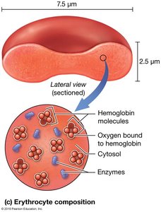
Erythrocytes are highly specialized cells responsible for gas transport. They are biconcave discs, which increases their surface area for rapid gas exchange and allows flexibility in capillaries.
Hematocrit: The percentage of blood volume occupied by RBCs; average is 46% in males and 42% in females.
Function: Transport oxygen and carbon dioxide via hemoglobin.
Specialization: Mature RBCs lack most organelles, rely on anaerobic glycolysis, and contain abundant hemoglobin.
Hemoglobin: Structure and Gas Exchange
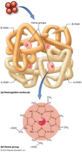
Hemoglobin is a quaternary globular protein with four subunits, each containing a heme group with iron. It binds oxygen reversibly at the iron center and carbon dioxide at the globin chains.
Oxyhemoglobin: Hemoglobin bound to oxygen.
Carbaminohemoglobin: Hemoglobin bound to carbon dioxide.
Deoxyhemoglobin: Hemoglobin not bound to oxygen.
Gas Exchange Basics
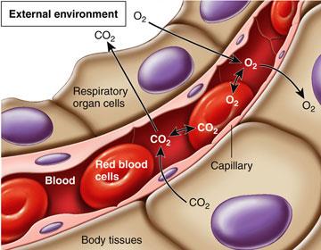
Oxygen binds to hemoglobin in the lungs and is released in peripheral tissues. Blood appears bright red when oxygenated and dull red when deoxygenated.
Fetal Hemoglobin and Other Variants
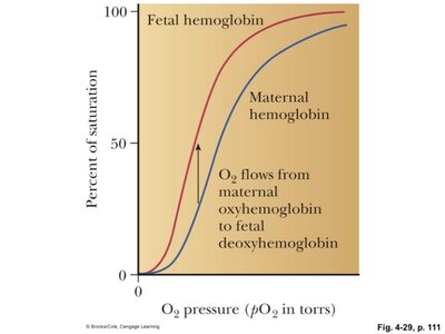
Fetal hemoglobin (HbF) has a higher affinity for oxygen than adult hemoglobin, allowing efficient oxygen transfer from mother to fetus. Other molecules, such as carbon monoxide, can compete for hemoglobin binding sites, reducing oxygen transport.
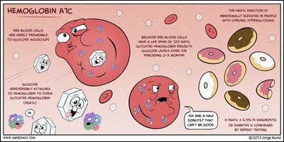
Glycated Hemoglobin (HbA1c)
HbA1c reflects average blood glucose concentration over the previous three months and is used as a diagnostic criterion for diabetes mellitus.
Erythrocyte Formation and Homeostasis
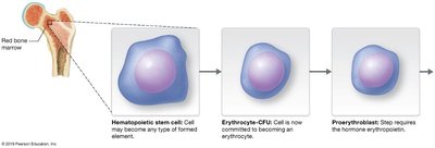
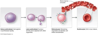
Erythropoiesis
Erythropoiesis is the process of RBC formation, occurring in myeloid tissue and requiring amino acids, iron, vitamin B12, and folic acid. The process involves several stages, from hematopoietic stem cells to mature erythrocytes.
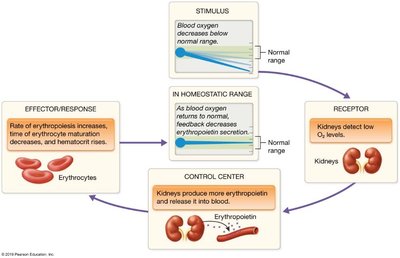
Regulation of Erythrocyte Production
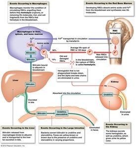
Erythrocyte homeostasis is maintained by a negative feedback loop. Tissue hypoxia stimulates the kidneys to release erythropoietin (EPO), increasing RBC production.
Erythrocyte Turnover and Disorders
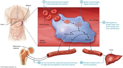
Turnover and Recycling
Damaged erythrocytes are phagocytized by macrophages in the liver, spleen, and bone marrow. Hemoglobin is broken down, iron is recycled, and heme is converted to bilirubin for excretion.
Anemias
Anemia is a reduction in the oxygen-carrying capacity of blood, caused by impaired RBC production, decreased hemoglobin, or abnormal hemoglobin. Symptoms include pallor, fatigue, weakness, and cardiac dysfunction.
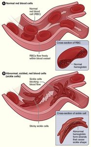
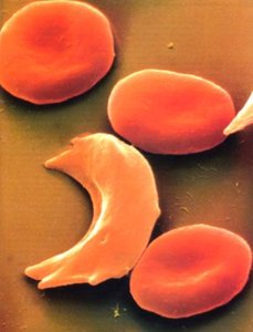
Sickle Cell Disease
Sickle cell disease is an inherited disorder caused by a mutation in the β-globin gene, resulting in abnormal hemoglobin (Hb-S) and sickled RBCs. These cells are prone to rupture, leading to hemolytic anemia and painful crises.
Formed Elements: Leukocytes (White Blood Cells)
Leukocyte Functions and Characteristics
Leukocytes are complete cells with nuclei and organelles, accounting for less than 1% of blood volume. They defend the body against pathogens and can move out of the bloodstream via diapedesis.
Leukopenia: Deficiency in leukocytes.
Leukocytosis: Excess leukocytes, often during infection.
Leukemia: Cancer of white blood cells, characterized by extreme leukocytosis.
Leukopoiesis
Leukopoiesis is the formation of WBCs, driven by hormones called interleukins and colony stimulating factors. Lymphocyte production is primarily antigen-driven.
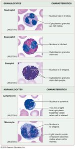
Types of Leukocytes
Type | Characteristics | Functions |
|---|---|---|
Neutrophils | 50–70% of WBCs; 3–5 lobed nucleus; granulocyte | Phagocytosis of bacteria; release enzymes and pro-inflammatory mediators |
Lymphocytes | 30% of WBCs; large nucleus; agranulocyte | T cells (cell-mediated immunity), B cells (antibody production), NK cells (immune surveillance) |
Monocytes | 4–8% of WBCs; largest; U-shaped nucleus; agranulocyte | Differentiate into macrophages; phagocytosis |
Eosinophils | 2–4% of WBCs; bi-lobed nucleus; granulocyte | Defense against parasites; phagocytosis of antibody-coated particles; inflammation |
Basophils | 1% of WBCs; U/S-shaped nucleus; granulocyte | Release histamine and heparin; allergic response |
Formed Elements: Thrombocytes (Platelets)
Platelet Structure and Function
Platelets are cell fragments from megakaryocytes, circulating for 7–10 days. They form temporary plugs, secrete vasoconstrictors and clotting factors, and contract after clot formation.
Hemostasis: Blood Clotting Mechanisms
Phases of Hemostasis
Hemostasis is the process that limits blood loss after trauma, involving five phases:
Vascular phase
Platelet phase
Coagulation phase
Clot retraction phase
Thrombolysis
Coagulation Pathways
Coagulation involves intrinsic and extrinsic pathways, both leading to the activation of Factor X and the formation of a fibrin clot. Calcium ions and vitamin K are essential for clotting factor synthesis.
Blood Types and Transfusion Compatibility
ABO and Rh Blood Groups
Blood type is determined by the presence of A, B, and D (Rh) antigens on erythrocytes. Compatibility is crucial for transfusions to prevent agglutination and hemolysis.
Type A: A antigens, anti-B antibodies
Type B: B antigens, anti-A antibodies
Type AB: Both antigens, no antibodies
Type O: No antigens, both antibodies
Rh factor: + (D antigen present), - (D antigen absent)
Clinical Perspectives and Disorders
Anemias
Iron-deficiency anemia: Microcytic, hypochromic RBCs; caused by blood loss or inadequate iron intake.
Pernicious anemia: Macrocytic, normochromic RBCs; caused by impaired vitamin B12 absorption.
Polycythemia vera: Overproduction of RBCs; increases blood viscosity and risk of vessel occlusion.
Leukemia
Leukemia is classified as acute or chronic, and lymphocytic or myelocytic, depending on the affected cell lineage and progression rate. It leads to anemia, thrombocytopenia, and increased immature WBCs.
Exercise and Blood Volume
Adaptations to Exercise
Chronic exercise increases blood volume, primarily through plasma expansion, followed by erythrocyte volume increase. This adaptation enhances oxygen delivery and aerobic capacity (VO2max).
*Additional info: Academic context was added to clarify the structure and function of blood components, hemoglobin variants, and clinical disorders. Tables and images were selected to directly reinforce the explanations provided.*
