 Back
BackChapter 19: Blood – Structure, Function, and Hemostasis
Study Guide - Smart Notes
 Tailored notes based on your materials, expanded with key definitions, examples, and context.
Blood: Introduction and Functions
Overview of Blood
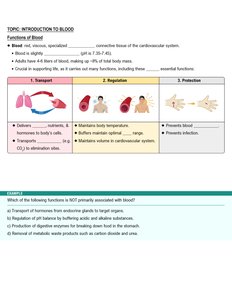
Blood is a specialized connective tissue that plays a vital role in the cardiovascular system. It is a red, viscous fluid that circulates throughout the body, delivering essential substances and removing waste products. Adults typically have 4–6 liters of blood, accounting for approximately 8% of total body mass.
Blood is slightly alkaline (pH 7.35–7.45).
It is crucial for supporting life by performing several essential functions.
Functions of Blood
Transport: Delivers oxygen and nutrients to body cells, removes metabolic wastes, and transports hormones.
Regulation: Maintains body temperature, stabilizes pH, and regulates fluid volume.
Protection: Prevents infection and blood loss through clotting mechanisms.
Composition of Blood
Main Components
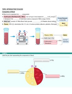
Blood consists of two main components: plasma (the liquid matrix) and formed elements (cells and cell fragments).
Erythrocytes (Red Blood Cells, RBCs): Transport oxygen and carbon dioxide; make up about 44% of blood volume.
Leukocytes (White Blood Cells, WBCs): Defend against pathogens.
Platelets (Thrombocytes): Involved in blood clotting.
Plasma: Straw-colored fluid (about 55% of blood) containing water, electrolytes, proteins (albumin, globulins, fibrinogen), nutrients, hormones, and waste products.
Erythrocytes (Red Blood Cells)
Structure of Erythrocytes
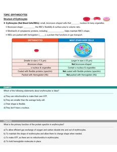
Erythrocytes are small, biconcave-shaped cells that lack nuclei and most organelles. Their shape increases surface area for gas exchange and allows flexibility to pass through capillaries.
Biconcave shape: Maximizes surface area-to-volume ratio for efficient gas exchange.
Lack of organelles: More room for hemoglobin, the protein responsible for oxygen transport.
Function of Hemoglobin
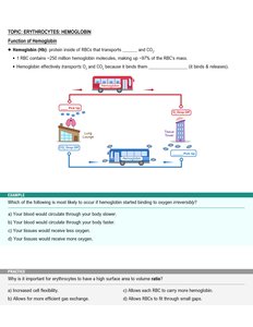
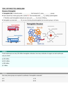
Hemoglobin (Hb) is the protein inside RBCs that binds and transports oxygen (O2) and carbon dioxide (CO2).
Each RBC contains about 250 million hemoglobin molecules.
Hemoglobin binds O2 in the lungs and releases it in tissues; it also transports some CO2 from tissues to lungs.
Structure of Hemoglobin
Hemoglobin is a tetrameric protein composed of four polypeptide chains (two alpha and two beta), each with a heme group that contains iron. Each heme binds one O2 molecule, so one hemoglobin can carry four O2 molecules.
Heme group: Iron-containing structure that binds oxygen reversibly.
Globin: Protein portion made of four polypeptide chains.
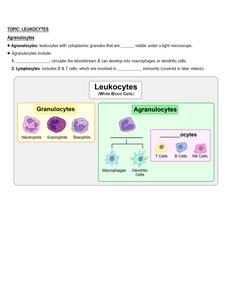
Leukocytes (White Blood Cells)
Introduction to Leukocytes
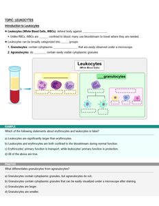
Leukocytes are immune cells that defend the body against infection and foreign substances. Unlike RBCs, WBCs have nuclei and can leave the bloodstream to reach tissues.
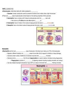
Granulocytes: Contain visible cytoplasmic granules (neutrophils, eosinophils, basophils).
Agranulocytes: Lack visible granules (lymphocytes, monocytes).
Granulocytes
Neutrophils: Most abundant WBC; phagocytize bacteria and are first responders to infection.
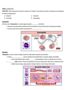
Eosinophils: Combat parasitic infections and are involved in allergic responses.
Basophils: Release histamine during allergic reactions and inflammation.
Agranulocytes
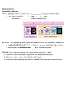
Lymphocytes: Include B cells (produce antibodies), T cells (cell-mediated immunity), and natural killer (NK) cells (destroy abnormal cells).
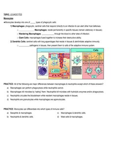
Monocytes: Differentiate into macrophages and dendritic cells, which phagocytize pathogens and present antigens to lymphocytes.
Platelets and Hemostasis
Introduction to Platelets
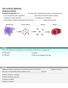
Platelets (thrombocytes) are cell fragments derived from megakaryocytes. They play a crucial role in stopping blood loss by forming plugs in damaged vessels and initiating clotting.
Structure: Lack nuclei, contain granules with clotting factors.
Function: Adhere to damaged vessel walls, aggregate, and release chemicals to promote clotting.
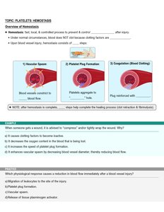
Hemostasis: The Process of Blood Clotting
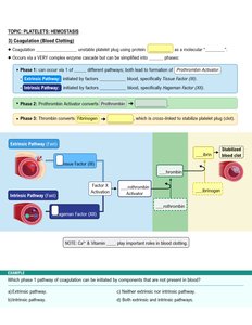
Hemostasis is the process that stops bleeding after vessel injury. It involves three main steps:
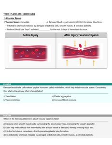
Vascular Spasm: Vasoconstriction reduces blood flow to the injured area.
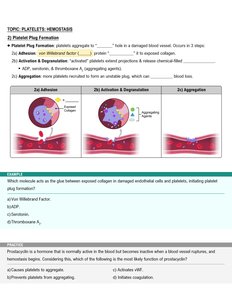
Platelet Plug Formation: Platelets adhere to exposed collagen and aggregate to form a temporary plug.
Coagulation (Blood Clotting): A cascade of reactions converts fibrinogen to fibrin, stabilizing the platelet plug.
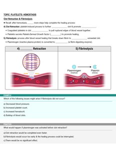
Clot Retraction and Fibrinolysis
After a clot forms, it retracts to bring wound edges together. Fibrinolysis then dissolves the clot once healing is underway.
Clot Retraction: Platelets contract, pulling fibrin threads together and tightening the clot.
Fibrinolysis: Plasminogen is converted to plasmin, which digests fibrin and dissolves the clot.
Summary Table: Main Formed Elements of Blood
Formed Element | Main Function | Relative Abundance |
|---|---|---|
Erythrocytes (RBCs) | Transport O2 and CO2 | ~99% of formed elements |
Leukocytes (WBCs) | Immune defense | ~1% of formed elements |
Platelets | Blood clotting | ~1% of formed elements |
Key Equations and Concepts
Oxygen Carrying Capacity: Each hemoglobin molecule binds 4 O2 molecules. Each RBC contains about 250 million hemoglobin molecules.
Hematocrit: Percentage of blood volume occupied by RBCs. Normal range: 36–45% (varies by sex and age).
O2 molecules
Additional info: These notes are based on Amerman, Human Anatomy & Physiology, Ch. 19 Blood, and are structured to provide a comprehensive overview suitable for ANP college students preparing for exams.
