 Back
Chapter 19: Blood – Structure, Function, and Clinical Relevance
Study Guide - Smart Notes
 Tailored notes based on your materials, expanded with key definitions, examples, and context.
Blood: An Introduction to the Cardiovascular System
Overview of the Cardiovascular System
The cardiovascular system consists of the heart, blood, and blood vessels. Blood is a specialized fluid connective tissue that transports nutrients, hormones, oxygen, and waste products throughout the body. It also plays a critical role in immune defense and homeostasis.
Transportation: Delivers gases, nutrients, hormones, and metabolic wastes.
Regulation: Maintains pH, ion composition, and body temperature.
Protection: Restricts fluid loss, defends against toxins and pathogens.
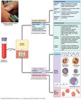
Blood: Functions and Physical Characteristics
Physical Properties of Blood
Blood is slightly denser than water, with a viscosity about five times greater. It is slightly alkaline (pH ~7.4) and its volume varies by sex (men: 5–6 L, women: 4–5 L).
Whole blood: Composed of plasma (fluid matrix) and formed elements (cells and cell fragments).
Formed elements: Include red blood cells (RBCs), white blood cells (WBCs), and platelets.
Hemopoiesis (hematopoiesis): The process of blood cell production.
Blood Plasma
Composition and Plasma Proteins
Plasma makes up 46–63% of whole blood and is about 92% water. It contains dissolved proteins, nutrients, hormones, and waste products. Plasma proteins are primarily synthesized by the liver and include:
Albumins (60%): Maintain osmotic pressure and transport fatty acids, hormones, and steroids.
Globulins (35%): Include immunoglobulins (antibodies) and transport globulins for hormones, metals, and lipids.
Fibrinogen (4%): Essential for blood clotting; converted to fibrin during coagulation.
Other proteins (<1%) include hormones such as insulin, prolactin, and TSH.
Red Blood Cells (RBCs)
Structure and Function
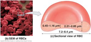
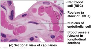
RBCs (erythrocytes) constitute 99.9% of formed elements and are responsible for oxygen transport. Their biconcave disc shape increases surface area for gas exchange and allows flexibility in capillaries.
Hematocrit: Percentage of blood volume occupied by RBCs; higher in males due to androgen stimulation.
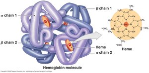
Hemoglobin: The oxygen-carrying protein in RBCs, composed of two alpha and two beta chains, each with a heme group containing iron.
RBC Life Cycle and Breakdown
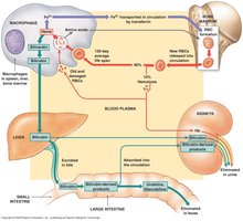
RBCs lack nuclei and most organelles, relying on anaerobic glycolysis for energy. They are removed from circulation by macrophages in the liver, spleen, and bone marrow before they rupture (hemolyze).
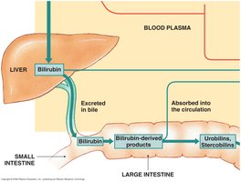
Hemoglobin breakdown: Heme is converted to biliverdin (green), then to bilirubin (yellow-orange), which is transported to the liver and excreted in bile.
Iron recycling: Iron is stored as ferritin or hemosiderin or transported by transferrin for reuse in erythropoiesis.
Clinical relevance: Excess bilirubin causes jaundice; hemoglobinuria and hematuria indicate kidney damage.
RBC Production (Erythropoiesis)
Sites and Stages of Erythropoiesis
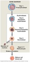
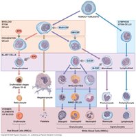
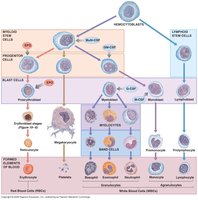
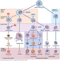
Erythropoiesis occurs in red bone marrow in adults. Hemocytoblasts (multipotent stem cells) differentiate into myeloid stem cells, which give rise to RBCs through several stages: proerythroblast, erythroblast, normoblast, reticulocyte, and mature RBC.
Regulation of Erythropoiesis
Erythropoiesis requires adequate supplies of amino acids, iron, and vitamins (B12, B6, folic acid). Erythropoietin (EPO), produced by the kidneys and liver in response to hypoxia, stimulates RBC production. Blood doping involves artificially increasing EPO to enhance athletic performance.
RBC Tests and Terminology
Common Laboratory Tests
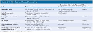
Several tests assess RBC function and health. Abnormal values can indicate anemia, polycythemia, or other disorders.
Test | Determines | Elevated | Suppressed |
|---|---|---|---|
Hematocrit (Hct) | % of formed elements in whole blood | Polycythemia | Anemia |
Reticulocyte count | % of circulating reticulocytes | Erythropoiesis/reticulocytosis | Anemia |
Hemoglobin concentration | g/dL of whole blood | Polycythemia | Anemia |
RBC count | Number per μL of blood | Erythrocytosis | Anemia |
Mean corpuscular volume (MCV) | Average volume of single RBC | Macrocytic | Microcytic |
Mean corpuscular hemoglobin concentration (MCHC) | Average Hb concentration in RBCs | Hyperchromic | Hypochromic |
Blood Types and Transfusion Compatibility
ABO and Rh Blood Groups
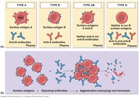
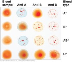
Blood types are determined by the presence of specific antigens (agglutinogens) on RBC membranes. The ABO system includes types A, B, AB, and O, while the Rh system classifies blood as Rh+ or Rh-.
Antibodies (agglutinins): Present in plasma, attack foreign antigens, causing agglutination (clumping) and hemolysis.
Type O-: Universal donor; Type AB+: Universal recipient.
Hemolytic Disease of the Newborn (HDN)
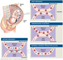
HDN occurs when maternal antibodies attack fetal RBCs, most commonly due to Rh incompatibility. It can be prevented by administering RhoGam to Rh-negative mothers during pregnancy.
White Blood Cells (WBCs)
Types and Functions of Leukocytes
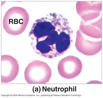
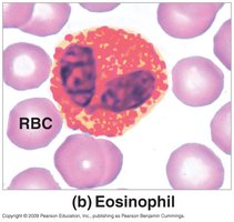
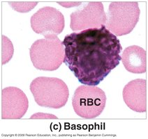
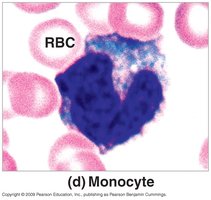
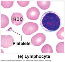
WBCs (leukocytes) are nucleated cells involved in immune defense. They can migrate out of the bloodstream, move via amoeboid motion, respond to chemical signals (chemotaxis), and some are phagocytic.
Nonspecific defense: Neutrophils, eosinophils, basophils, monocytes.
Specific defense: Lymphocytes (T cells, B cells, NK cells).
WBC Production and Regulation
WBCs are produced from hemocytoblasts in the bone marrow. Myeloid stem cells give rise to all WBCs except lymphocytes, which derive from lymphoid stem cells. Colony-stimulating factors (CSFs) regulate the production of specific WBC types.
Platelets and Hemostasis
Platelet Structure and Function
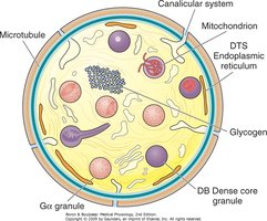
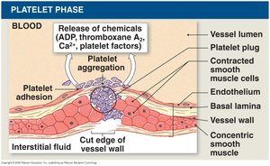
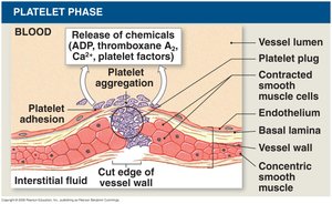
Platelets are cell fragments derived from megakaryocytes and are essential for blood clotting. They release clotting chemicals, form temporary plugs, and contract to reduce clot size.
Hemostasis: The Process of Blood Clotting
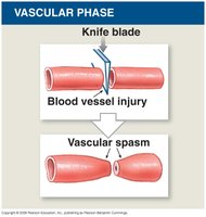
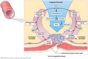
Hemostasis is the cessation of bleeding and involves three phases:
Vascular phase: Vasoconstriction and endothelial changes at the injury site.
Platelet phase: Platelet adhesion, aggregation, and plug formation.
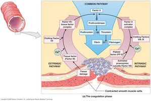
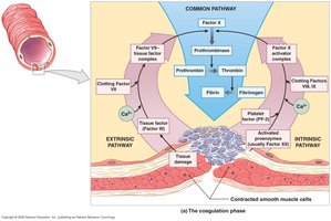
Coagulation phase: Cascade of clotting factors leading to fibrin formation.
Feedback and Regulation of Clotting
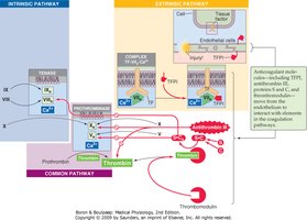
Anticoagulants such as antithrombin-III, heparin, and protein C regulate clot formation. Prostacyclin inhibits platelet aggregation. Fibrinolysis, the dissolution of clots, is mediated by plasmin generated from plasminogen.
Role of Calcium Ions and Vitamin K
Calcium ions (Ca2+) are required for all steps of the clotting cascade. Vitamin K is necessary for the synthesis of several clotting factors by the liver. Deficiencies in either can impair hemostasis.
Additional info: Vitamin K is fat-soluble and can be obtained from diet or synthesized by intestinal bacteria. Broad-spectrum antibiotics or fat malabsorption can lead to clotting disorders due to vitamin K deficiency.
