 Back
BackChapter 19: Blood Vessels – Structure, Function, and Circulation
Study Guide - Smart Notes
 Tailored notes based on your materials, expanded with key definitions, examples, and context.
Blood Vessels: Overview
Introduction to Blood Vessels
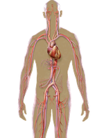
Blood vessels are essential components of the cardiovascular system, responsible for transporting blood throughout the body. They include arteries, veins, and capillaries, each with distinct structural and functional characteristics.
Arteries: Carry blood away from the heart.
Veins: Carry blood toward the heart.
Capillaries: Connect arteries and veins; site of nutrient and waste exchange.
Gross and Microscopic Anatomy of Blood Vessels
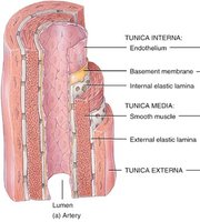
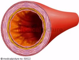
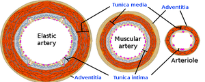
Layers of Arteries and Veins
Blood vessels are composed of three main layers, or tunics, which vary in thickness and composition between arteries and veins.
Tunica Intima: Innermost layer; consists of endothelium (simple squamous epithelium) and basement membrane. Provides a smooth lining for blood flow.
Tunica Media: Middle layer; composed of smooth muscle and elastic fibers. Responsible for vasoconstriction and vasodilation, regulated by the sympathetic nervous system.
Tunica Externa (Adventitia): Outermost layer; made of connective tissue with collagen and elastic fibers. Provides structural support and protection.

Classification of Arteries
Types of Arteries
Arteries are classified based on their size and function:
Elastic Arteries (Conduction Arteries): Largest arteries near the heart (e.g., aorta). Contain abundant elastic fibers, allowing them to act as pressure reservoirs and maintain continuous blood flow.
Muscular Arteries: Medium-sized arteries with more smooth muscle than elastic fibers. Deliver blood to specific body regions (e.g., brachial, femoral arteries).
Arterioles: Smallest arteries; heavily innervated and regulate blood flow to capillary beds through vasoconstriction and vasodilation.

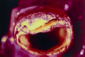
Arteriosclerosis and Atherosclerosis
Definitions and Pathology
Arteriosclerosis: General term for degenerative changes in arteries leading to decreased elasticity.
Atherosclerosis: Specific form of arteriosclerosis characterized by lipid deposits (plaques) in large arteries, leading to reduced blood flow and increased risk of cardiovascular events.
Capillaries: Structure and Function
Capillary Anatomy and Classification
Capillaries are the smallest blood vessels, consisting only of tunica intima (endothelium and basement membrane). They provide direct access to nearly every cell in the body and are the primary site for nutrient and waste exchange.
Continuous Capillaries: Most common; tight junctions with intercellular clefts allow passage of fluids and small solutes. Found in skin, muscles, and brain (blood-brain barrier).
Fenestrated Capillaries: Endothelial cells have pores; allow filtration and are found in kidneys, small intestine, and endocrine glands.
Sinusoidal Capillaries: Large, irregular lumen; incomplete basement membrane and large gaps. Highly permeable, found in liver, bone marrow, and spleen.
Capillary Beds and Blood Flow Regulation
Structure of Capillary Beds
Capillary beds consist of a network of capillaries supplied by arterioles and drained by venules.
Metarteriole (Vascular Shunt): Connects arteriole to venule; acts as a thoroughfare channel.
True Capillaries: Branch from the metarteriole; actual sites of exchange.
Precapillary Sphincters: Rings of smooth muscle that regulate blood flow into true capillaries based on local chemical conditions and neural input.
Veins: Structure and Function
Characteristics of Veins
Veins have thinner walls, larger lumens, and lower blood pressure compared to arteries. They act as blood reservoirs, containing up to 65% of the body's blood supply.
Tunica Intima: Forms valves to prevent backflow.
Tunica Media: Thinner than in arteries.
Tunica Externa: Thickest layer in veins.
Vascular Anastomoses
Interconnections of Blood Vessels
Arterial Anastomoses: Provide alternate pathways (collateral channels) for blood flow, especially important in joints, abdominal organs, brain, and heart.
Venous Anastomoses: More common; allow for multiple routes of venous return.
Blood Flow and Blood Pressure
Definitions and Measurement
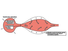
Blood Flow: Volume of blood flowing through a tissue per unit time (mL/min). Total blood flow equals cardiac output.
Blood Pressure: Force per unit area exerted on vessel walls. Highest in the aorta, lowest in the venae cavae.
Systolic Pressure: Pressure during ventricular contraction (~120 mmHg).
Diastolic Pressure: Pressure during ventricular relaxation (~70 mmHg).
Pulse Pressure: Difference between systolic and diastolic pressure.
Mean Arterial Pressure (MAP): Average pressure during a cardiac cycle.
Factors Influencing Blood Pressure
Peripheral Resistance, Blood Volume, and Cardiac Output
Peripheral Resistance: Opposition to flow; affected by blood viscosity, vessel length, and vessel diameter.
Blood Volume: Regulated by kidneys; affects venous return and cardiac output.
Cardiac Output: (Stroke Volume × Heart Rate)
Regulation of Blood Pressure
Neural and Hormonal Control
Short-term Regulation: Neural (baroreceptors, chemoreceptors) and hormonal (epinephrine, norepinephrine, angiotensin II, ADH, ANP) mechanisms alter vessel diameter and heart rate.
Long-term Regulation: Kidneys adjust blood volume via direct and indirect mechanisms (renin-angiotensin-aldosterone system).
Blood Pressure Disorders
Hypertension and Hypotension
Hypertension: Sustained arterial pressure of 140/90 mmHg or higher. Risk factors include heredity, diet, obesity, age, diabetes, stress, and smoking.
Hypotension: Blood pressure below 90/60 mmHg. May be orthostatic, acute (shock), or chronic (Addison’s disease, hypothyroidism).
Capillary Exchange Mechanisms
Diffusion and Bulk Flow
Diffusion: Movement of molecules along concentration gradients through intercellular clefts, fenestrations, or cell membranes.
Bulk Flow: Movement of fluid and solutes due to pressure gradients.
Hydrostatic Pressure (HP): Pushes fluid out of capillaries.
Osmotic Pressure (OP): Draws fluid into capillaries.
Circulatory Shock
Types and Causes
Hypovolemic Shock: Due to large-scale fluid loss.
Vascular Shock: Due to extreme vasodilation and loss of vasomotor tone.
Cardiogenic Shock: Due to heart failure or damage.
Circulatory Pathways
Systemic, Pulmonary, and Special Circulations
Systemic Circulation: Delivers oxygen and nutrients to tissues; removes wastes.
Pulmonary Circulation: Delivers deoxygenated blood to lungs for gas exchange.
Hepatic Portal Circulation: Shunts nutrient-rich blood from GI tract to liver for processing.
Summary Table: Types of Blood Vessels
Type | Structure | Function |
|---|---|---|
Artery | Thick tunica media, elastic fibers | Carry blood away from heart |
Vein | Thin walls, large lumen, valves | Carry blood toward heart |
Capillary | Single layer endothelium | Site of exchange |
Summary Table: Capillary Types
Type | Structure | Location | Permeability |
|---|---|---|---|
Continuous | Tight junctions, intercellular clefts | Skin, muscles, brain | Low |
Fenestrated | Pores in endothelium | Kidneys, intestines, glands | Medium |
Sinusoidal | Large gaps, incomplete membrane | Liver, bone marrow, spleen | High |
