 Back
BackChapter 19: The Cardiovascular System – Blood Vessels (Mini-Textbook Study Guide)
Study Guide - Smart Notes
 Tailored notes based on your materials, expanded with key definitions, examples, and context.
Blood Vessel Structure and Function
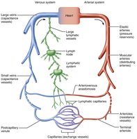
Overview of Blood Vessels
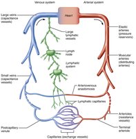
Blood vessels form a closed delivery system that begins and ends at the heart, working dynamically to pulsate, constrict, relax, and multiply. They collaborate with the lymphatic system to circulate fluids throughout the body.
Veins carry blood toward the heart. Systemic veins carry oxygen-poor blood, while pulmonary veins carry oxygenated blood. Umbilical veins transport oxygenated blood from placenta to fetus.
Arteries carry blood away from the heart. Systemic arteries carry oxygenated blood, pulmonary arteries carry oxygen-poor blood, and umbilical arteries transport oxygen-poor blood from fetus to placenta.
Capillaries are exchange vessels, directly serving cellular needs by allowing substances to move across their walls between tissue cells and blood.
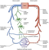
Layers of Blood Vessel Walls
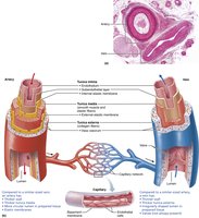
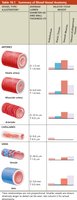
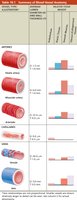
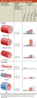
Most blood vessel walls (except capillaries) consist of three layers, or tunics:
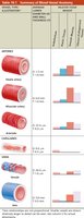
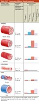
Tunica intima: Innermost layer, in intimate contact with blood. Composed of endothelium (simple squamous epithelium) and a subendothelial layer (basement membrane and loose connective tissue).
Tunica media: Middle layer, mostly circularly arranged smooth muscle cells. Responsible for vasoconstriction (decreasing lumen diameter) and vasodilation (increasing lumen diameter), thus regulating blood flow and pressure.
Tunica externa (adventitia): Outermost layer, composed mostly of collagen fibers that protect, reinforce, and anchor the vessel. Contains nerve fibers, lymphatic vessels, and blood vessels (vasa vasorum).
Capillary walls consist only of endothelium with a sparse basal lamina. 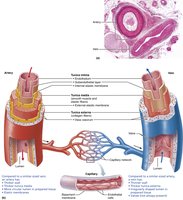
Vasa Vasorum
The vasa vasorum are small blood vessels found in the walls of larger vessels, nourishing the external tissues of the vessel wall. 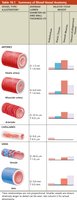
Arteries: Types and Functions
Classification of Arteries
Arteries are divided into three groups based on size and function:
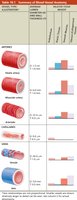
Elastic arteries: Thick-walled arteries near the heart (e.g., aorta, pulmonary trunk). Act as pressure reservoirs, expanding and recoiling to maintain continuous blood flow.
Muscular arteries: Distributing arteries that deliver blood to specific organs. Have the thickest tunica media and are more vasoactive (responsive to diameter changes).
Arterioles: Smallest arteries, leading into capillary beds. Control flow into capillaries via vasodilation and vasoconstriction, and are major determinants of total peripheral resistance (TPR).
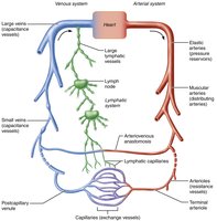
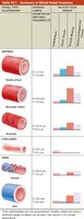
Elastic Arteries
Large lumen provides low resistance to blood flow.
Contain more elastin than other vessels, found in all three tunics.
Act as pressure reservoirs, expanding and recoiling as blood is ejected from the heart.
Diseases like atherosclerosis can stiffen these arteries, leading to aneurysms or rupture.
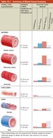
Muscular Arteries
Deliver blood to specific body organs.
Have thickest tunica media, more smooth muscle, less elastic tissue.
Less stretchy, more vasoactive than elastic arteries.
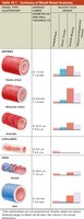
Arterioles
Smallest arteries, leading into capillary beds.
Larger arterioles have all three tunics; smaller ones are mostly smooth muscle and endothelium.
Control flow into capillary beds and resistance to blood flow.
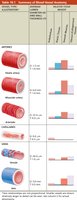
Capillaries: Exchange Vessels
Structure and Function
Capillaries are the smallest blood vessels, consisting only of thin tunica intima. Their ultra-thin walls allow for exchange of materials (gases, nutrients, wastes, hormones) between blood and interstitial fluid.
Most tissues have a rich capillary supply, except poorly vascularized tendons, ligaments, cartilage, epithelia, cornea, and lens.
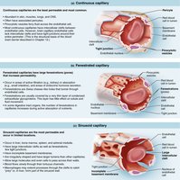
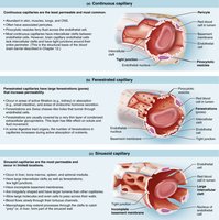
Some capillaries have pericytes (contractile stem cells) that stabilize the wall and control permeability.
Types of Capillaries
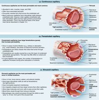
Continuous capillaries: Least permeable, most common. Abundant in skin, muscles, lungs, and CNS.
Fenestrated capillaries: Found in areas of active filtration (kidneys), absorption (intestines), and endocrine hormone secretion. Have pores (fenestrations) for increased permeability.
Sinusoidal capillaries: Most permeable, least common. Found in liver, bone marrow, spleen, and adrenal medulla. Have large clefts, fenestrations, and incomplete basement membranes.
Capillary Beds
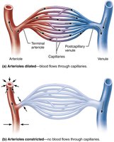
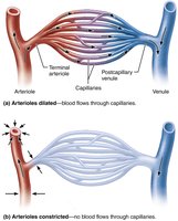
Capillary beds are interwoven networks between arterioles and venules, providing microcirculation. Blood flow through the bed is controlled by the diameter of terminal arterioles and upstream arterioles, regulated by chemicals and nerve fibers.
Dilating arterioles increases blood flow; constricting decreases it.
Blood flow matches tissue metabolic demands.
Veins: Blood Reservoirs
Structure and Function
Veins carry blood away from capillary beds toward the heart. As venous vessels join, they form larger diameters and thicker walls.
Veins have all tunics, but thinner walls and larger lumens compared to arteries.
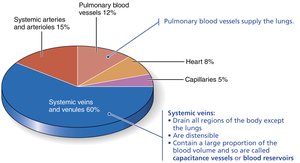
Contain up to 65% of blood supply at any time, acting as capacitance vessels and blood reservoirs.
Blood pressure is lower than in arteries; adaptations ensure return of blood to heart.
Venules
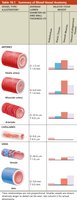
Formed by uniting capillaries; smallest venules consist only of endothelium and pericytes.
Very porous, allowing fluids and WBCs to move into tissues.
Larger venules have thin tunica media and externa.
Veins
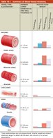
Thinner walls, larger lumens, collapsed appearance in histology.
Tunica media is thin; tunica externa is much thicker.
Adaptations include large-diameter lumens (low resistance) and venous valves (prevent backflow).
Venous sinuses are specialized, flattened veins with thin walls, supported by surrounding tissues.
Blood Distribution
Systemic veins contain the most blood (about 60%).
Valves and large lumens help compensate for low venous pressure.
Blood Flow, Pressure, and Resistance
Definitions
Blood flow: Amount of blood flowing through a vessel, organ, or entire circulation per unit time (ml/min).
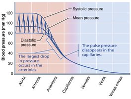
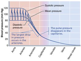
Blood pressure (BP): Force per unit area exerted on vessel wall by blood, measured in mm Hg.
Resistance: Opposition to flow, mainly due to friction along vessel walls. Measured as total peripheral resistance (TPR).
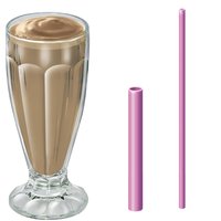
Three sources of resistance: blood viscosity, vessel length, and vessel diameter.
Relationship Between Flow, Pressure, and Resistance
Blood flow (F) is directly proportional to the pressure gradient (ΔP) and inversely proportional to TPR.
Equation:
Small-diameter arterioles are major determinants of TPR.
Resistance varies inversely with the fourth power of vessel radius:
Blood Pressure Regulation
Factors Affecting Blood Pressure
Cardiac output (CO), TPR, and blood volume are main factors.
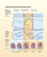
Anything increasing stroke volume (SV), heart rate (HR), or TPR increases mean arterial pressure (MAP).
MAP calculation:
Short-Term Regulation: Neural and Hormonal Controls
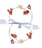
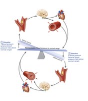
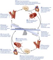
Neural controls maintain MAP by altering vessel diameter and blood distribution.
Baroreceptor reflexes monitor stretch in vessel walls and adjust MAP via the cardiovascular center in the medulla.
Chemoreceptor reflexes respond to changes in blood chemistry (O2, CO2, pH).
Hormones (epinephrine, norepinephrine, ADH, ANP) regulate MAP by affecting CO, TPR, and blood volume.
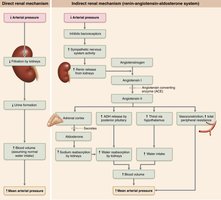
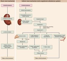
Long-Term Regulation: Renal Mechanisms
Kidneys regulate blood volume and MAP via direct and indirect mechanisms.
Direct mechanism: Kidneys eliminate or conserve water based on blood pressure.
Indirect mechanism: Renin-angiotensin-aldosterone system increases MAP by vasoconstriction, water retention, and stimulating thirst.
Blood Flow Regulation
Intrinsic and Extrinsic Controls
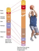
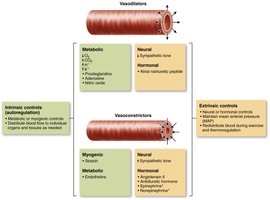
Intrinsic controls (autoregulation): Organs regulate their own blood flow by varying resistance of their arterioles.
Extrinsic controls: Sympathetic and endocrine mechanisms maintain MAP and redistribute blood flow as needed.
During exercise, intrinsic controls increase blood flow to muscles, while extrinsic controls decrease flow to kidneys and digestive organs.
Capillary Exchange
Fluid Compartments and Exchange
Two major fluid compartments: extracellular fluid (plasma and interstitial fluid) and intracellular fluid.
Exchange of gases, nutrients, water, and wastes occurs between these compartments via capillary walls.
Velocity of Blood Flow
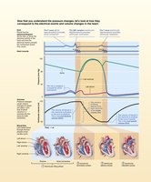
Velocity is fastest in the aorta, slowest in capillaries, and increases again in veins.
Speed is inversely related to total cross-sectional area; capillaries have the largest area, allowing slow flow for exchange.
Capillary Transport Mechanisms
Lipid-soluble molecules diffuse across endothelial cell membranes.
Water-soluble molecules pass through intercellular clefts or fenestrations.
Larger molecules are transported via endocytosis and transcytosis.
Bulk Flow: Hydrostatic and Osmotic Pressures
Fluid is forced out of capillaries at the arterial end (filtration) and returns at the venous end (reabsorption).
Hydrostatic pressure pushes fluid out; colloid osmotic pressure pulls fluid in.
Net filtration pressure (NFP) determines direction and amount of fluid movement:
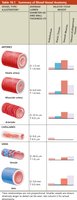
Summary Table: Blood Vessel Types
Type | Structure | Function | Key Features |
|---|---|---|---|
Elastic Arteries | Thick walls, large lumen, abundant elastin | Pressure reservoir | Expand/recoil, low resistance |
Muscular Arteries | Thick tunica media, more smooth muscle | Distribute blood to organs | Vasoactive, less elastic |
Arterioles | Smallest arteries, mostly smooth muscle | Control flow/resistance | Major TPR determinant |
Capillaries | Single layer endothelium | Exchange vessels | Permeability varies |
Veins | Thin walls, large lumen, valves | Return blood, reservoir | Low pressure, capacitance |
Key Equations
Blood flow:
Resistance:
Mean arterial pressure:
Net filtration pressure:
Additional info:
Baroreceptors and chemoreceptors are essential for short-term regulation of blood pressure.
Renin-angiotensin-aldosterone system is critical for long-term regulation via kidneys.
Capillary exchange is vital for tissue health and fluid balance.
