 Back
BackChapter 19: The Cardiovascular System – Blood
Study Guide - Smart Notes
 Tailored notes based on your materials, expanded with key definitions, examples, and context.
Cardiovascular System: Blood
Overview of Blood Functions
Blood is a specialized connective tissue that plays a vital role in the transport of substances, regulation of physiological parameters, and protection of the body. It is essential for maintaining homeostasis and supporting cellular activities throughout the body.
Distribution: Transports gases (O2, CO2), nutrients, wastes, and hormones to and from cells.
Regulation: Maintains body temperature, pH balance, and blood pressure.
Protection: Prevents blood loss (hemostasis) and provides immune defense against pathogens.
Physical Characteristics of Blood
Blood is an opaque, sticky fluid with a slightly alkaline pH and a higher viscosity than water. It constitutes about 8% of total body weight.
Volume: Males: 5–6 L; Females: 4–5 L
Temperature: Slightly higher than body temperature (about 38°C)
Viscosity: Determined by plasma proteins and formed elements
Blood Components
Blood consists of two major components: plasma and formed elements. Plasma is the liquid matrix, while formed elements include erythrocytes, leukocytes, and platelets.
Plasma: 55% of blood volume; contains water, proteins (albumin, globulins, fibrinogen), and solutes.
Formed Elements: 45% of blood volume; includes erythrocytes (RBCs), leukocytes (WBCs), and platelets (thrombocytes).
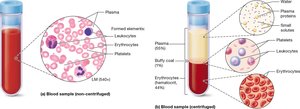
Hematopoiesis and Hematocrit
Hematopoiesis is the process of blood cell formation, primarily occurring in the red bone marrow. When blood is centrifuged, it separates into three layers:
Plasma: Top layer (~55%)
Buffy Coat: Middle layer (~1%), contains leukocytes and platelets
Erythrocytes: Bottom layer (~44%)
Hematocrit is the percentage of blood volume occupied by erythrocytes.
Plasma Composition
Plasma is a pale yellow fluid, making up about 55% of blood volume. It is composed of:
Water: 90% of plasma; solvent for carrying other substances
Plasma Proteins: 9%; includes albumin (osmotic balance), immune proteins (defense), transport proteins, and clotting proteins (fibrinogen)
Solutes: 1%; includes nutrients, electrolytes, gases, hormones, and metabolic wastes
Erythrocytes (Red Blood Cells)
Structure and Function
Erythrocytes are biconcave, anucleate cells specialized for oxygen and carbon dioxide transport. Their shape increases surface area for gas exchange and flexibility for passage through capillaries.
Main Function: Transport O2 (bound to hemoglobin) and CO2
Structure: Lack nucleus and most organelles; essentially sacs of hemoglobin
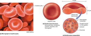
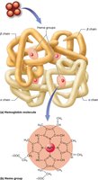
Hemoglobin
Hemoglobin (Hb) is the oxygen-carrying protein in erythrocytes. Each molecule consists of four subunits, each containing a heme group with an iron atom that binds O2.
Oxyhemoglobin: Hemoglobin bound to oxygen
Deoxyhemoglobin: Hemoglobin not bound to oxygen
CO2 Transport: CO2 binds to globin subunits
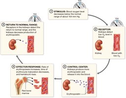
Erythrocyte Life Cycle and Erythropoiesis
Erythrocytes have a lifespan of about 120 days. Erythropoiesis is the process of erythrocyte production, stimulated by erythropoietin (EPO) from the kidneys in response to hypoxia (low oxygen levels).
Destruction: Old erythrocytes are removed by the spleen; components are recycled.
Heme Breakdown: Heme is converted to bilirubin and transported to the liver for excretion.
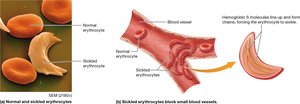
Erythrocyte Disorders
Anemia: Decreased oxygen-carrying capacity of blood. Causes include:
Decreased hemoglobin (iron-deficiency anemia)
Decreased hematocrit (hemolytic or aplastic anemia)
Abnormal hemoglobin (thalassemia, sickle cell disease)
Polycythemia: Excess erythrocytes, increasing blood viscosity and risk of clotting.
Leukocytes (White Blood Cells)
Structure and Function
Leukocytes are larger than erythrocytes, contain a nucleus, and are involved in immune defense. They can leave the bloodstream (diapedesis) to enter tissues.
Lifespan: 12–20 days
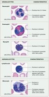
Types: Granulocytes and agranulocytes
Types of Leukocytes
Type | Main Function |
|---|---|
Neutrophil | Phagocytosis of bacteria; most numerous WBC |
Eosinophil | Active against parasitic worms and in allergic reactions |
Basophil | Release histamine; mediate inflammation |
Lymphocyte | Form T cells (destroy abnormal cells), B cells (produce antibodies), NK cells (immunological surveillance) |
Monocyte | Become macrophages; ingest dead cells, bacteria, debris |
Leukocyte Disorders
Leukemia: Cancer of blood-forming tissues; abnormal proliferation of WBCs.
Infectious Mononucleosis: Viral infection causing fever, fatigue, and swollen lymph nodes.
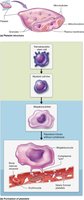
Platelets (Thrombocytes)
Structure and Function
Platelets are cell fragments derived from megakaryocytes. They play a crucial role in hemostasis (blood clotting) and have a lifespan of about 10 days.
Formation: Stimulated by thrombopoietin from the liver; megakaryocytes extend processes into capillaries, and fragments break off as platelets.
Function: Release clotting factors and form platelet plugs to stop bleeding.
Hemostasis
Overview of Hemostasis
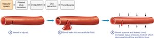
Hemostasis is the process that stops blood loss from damaged vessels. It involves a series of steps to form a stable blood clot.
1. Vascular spasm
2. Platelet plug formation
3. Coagulation (clotting cascade)
4. Clot retraction
5. Thrombolysis (clot removal)
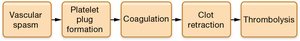
Step 1: Vascular Spasm
Vascular spasm is the immediate constriction of a damaged blood vessel to reduce blood flow and limit blood loss.
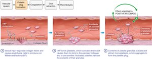
Step 2: Platelet Plug Formation
Platelets adhere to exposed collagen fibers at the injury site, become activated, and aggregate to form a temporary plug. This process is amplified by positive feedback.
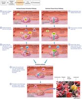
Step 3: Coagulation
Coagulation is a cascade of enzymatic reactions that convert fibrinogen to fibrin, forming a stable blood clot. It involves intrinsic and extrinsic pathways that converge on the activation of thrombin.
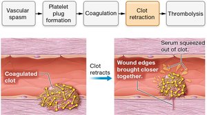
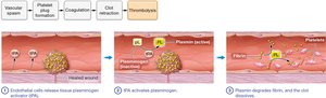
Steps 4 & 5: Clot Retraction and Thrombolysis
Clot retraction tightens the clot, bringing wound edges together. Thrombolysis dissolves the clot after tissue repair is complete.
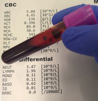
Clinical Assessment: Complete Blood Count (CBC)
A CBC is a common laboratory test that evaluates the number and characteristics of blood cells. It is used to diagnose anemia, infections, and other hematological disorders.
Parameters: RBC count, hematocrit, hemoglobin concentration, WBC count and differential, platelet count
Blood Disorders
Bleeding Disorders
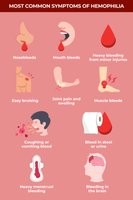
Hemophilia: Genetic disorder; deficiency of specific clotting factors leads to excessive bleeding.
Thrombocytopenia: Low platelet count; increased risk of bleeding.
Impaired Liver Function: Reduced synthesis of clotting factors.
Hypercoagulable Conditions
Thrombus: Clot forms in an unbroken blood vessel.
Embolus: A moving blood clot that can block vessels elsewhere in the body.
Blood Transfusions and Blood Typing
Blood transfusions involve transferring blood or blood components from a donor to a recipient. Blood typing (ABO and Rh systems) is essential to prevent transfusion reactions.
Types of Transfusions: Whole blood, packed RBCs, platelets, WBCs
Blood Typing: Determines compatibility for safe transfusion
Additional info: Blood typing is based on the presence or absence of antigens (A, B, Rh) on erythrocyte membranes. Incompatible transfusions can cause agglutination and hemolysis.
