 Back
BackChapter 19: The Circulatory System – Blood
Study Guide - Smart Notes
 Tailored notes based on your materials, expanded with key definitions, examples, and context.
The Circulatory System: Blood
Overview of Blood Circulation
Blood is a vital connective tissue responsible for transporting substances throughout the body. It circulates via arteries, capillaries, and veins, facilitating the exchange of gases, nutrients, and wastes between tissues and the bloodstream.
Arteries carry blood away from the heart, branching into capillaries.
Capillaries allow diffusion of oxygen and nutrients into tissues, and removal of carbon dioxide and wastes.
Veins return oxygen-deficient blood to the heart, which is then sent to the lungs for gas exchange.
Composition of Blood
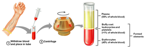
Blood consists of a liquid matrix called plasma and formed elements. It is the only fluid tissue in the body and is classified as connective tissue.
Plasma: Straw-colored fluid, ~90–92% water, ~8% proteins, ~1% other solutes.
Formed elements: Erythrocytes (RBCs), leukocytes (WBCs), and platelets.
Components of Whole Blood
Blood is separated into plasma and formed elements by centrifugation. The hematocrit is the percentage of RBCs in total blood volume.
Normal hematocrit: Males: 46%±5%, Females: 42%±5%
Physical Characteristics and Volume
Blood is a sticky, opaque fluid with a metallic taste. Its color varies from bright scarlet (oxygen-rich) to dark red (oxygen-poor).
pH: 7.35–7.45 (slightly alkaline)
Temperature: ~38°C (100.4°F)
Volume: Males: ~5–6 L, Females: ~4–5 L
Functions of Blood
Blood performs three main functions: distribution, regulation, and protection.
Distribution: Transports oxygen, nutrients, hormones, and wastes.
Regulation: Maintains pH, temperature, and fluid balance.
Protection: Hemostasis (clotting) and immune defense.
Blood Plasma
Plasma is the fluid matrix of blood, containing water, proteins, nutrients, electrolytes, gases, and hormones.
Proteins: Albumin (60%), globulins (35%), fibrinogen (4%)
Electrolytes: Sodium, potassium, calcium, chloride, bicarbonate
Organic nutrients: Glucose, amino acids, fatty acids, vitamins
Plasma Proteins
Plasma proteins are essential for osmotic balance, transport, and immune function.
Albumin: Maintains osmotic pressure, transports substances, buffers pH.
Globulins: Transport proteins (α, β) and antibodies (γ).
Fibrinogen: Essential for blood clotting.
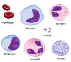
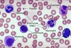
Formed Elements
Formed elements include erythrocytes, leukocytes, and platelets. Only WBCs are complete cells; RBCs lack nuclei, and platelets are cell fragments.
Lifespan: RBCs: ~120 days, Platelets: ~7–10 days, WBCs: hours to years
Origin: All formed elements arise from hematopoietic stem cells in red bone marrow.
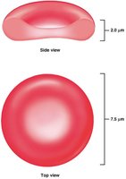
Erythrocytes (Red Blood Cells)
Structure and Function
Erythrocytes are small, biconcave, anucleate cells specialized for gas transport. Their shape increases surface area for efficient diffusion.
Diameter: ~7.5 μm
Hemoglobin: Major protein for oxygen and carbon dioxide transport
Flexibility: Allows passage through narrow capillaries
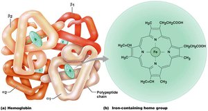
Hemoglobin Structure
Hemoglobin is a protein composed of four polypeptide chains (2 α, 2 β) and four heme groups, each containing an iron atom that binds oxygen.
Oxyhemoglobin: Hemoglobin bound to O₂ (bright red)
Deoxyhemoglobin: Hemoglobin after O₂ release (dark red)
Carbaminohemoglobin: Hemoglobin bound to CO₂
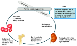
Production and Regulation of Erythrocytes
Erythrocyte production (erythropoiesis) occurs in red bone marrow and is regulated by erythropoietin (EPO), a hormone released by the kidneys in response to hypoxia.
Stem cell: Hemocytoblast differentiates into proerythroblast, then reticulocyte, then mature RBC.
Regulation: Negative feedback via EPO; increased RBCs suppress EPO production.
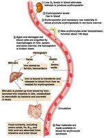
Life Cycle and Fate of Erythrocytes
RBCs have a lifespan of ~100–120 days. Old RBCs are removed by macrophages in the spleen, liver, and bone marrow. Hemoglobin is broken down and recycled.
Globin: Broken into amino acids
Heme: Iron recycled; remainder converted to bilirubin and excreted
Erythrocyte Disorders
Anemia is a condition of reduced oxygen-carrying capacity, caused by decreased RBC production, hemoglobin deficiency, or increased destruction.
Symptoms: Fatigue, pallor, shortness of breath, cold intolerance
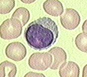
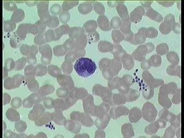
Leukocytes (White Blood Cells)
Types and Functions
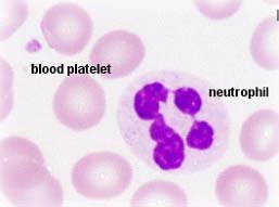
Leukocytes are complete cells essential for immune defense. They are classified as granulocytes or agranulocytes.
Granulocytes: Neutrophils, eosinophils, basophils
Agranulocytes: Lymphocytes, monocytes
Granulocytes
Granulocytes contain cytoplasmic granules and have lobed nuclei. They are involved in phagocytosis and inflammation.
Neutrophils: Most numerous, highly phagocytic, first responders to infection
Eosinophils: Target parasites and participate in allergic responses
Basophils: Release histamine and heparin, involved in inflammation
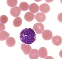
Agranulocytes
Agranulocytes lack visible granules. Lymphocytes are key to adaptive immunity, while monocytes differentiate into macrophages for phagocytosis.
Lymphocytes: B cells (antibody production), T cells (cell-mediated immunity), NK cells (destroy abnormal cells)
Monocytes: Largest WBCs, become macrophages in tissues
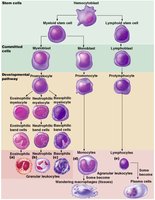
Leukocyte Production (Leukopoiesis)
Leukopoiesis is regulated by cytokines (interleukins and colony-stimulating factors) and occurs in bone marrow. All WBCs originate from hemocytoblasts.
Myeloid stem cells: Give rise to granulocytes and monocytes
Lymphoid stem cells: Give rise to lymphocytes
Platelets (Thrombocytes)
Structure and Function
Platelets are cell fragments essential for hemostasis. They form temporary plugs and initiate the clotting cascade at sites of vascular injury.
Lifespan: ~7–10 days
Granule contents: Serotonin, Ca²⁺, ADP, enzymes, PDGF
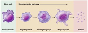
Platelet Formation (Thrombopoiesis)
Platelets are produced in the bone marrow from megakaryocytes, which fragment to release platelets into circulation.
Lineage: Hemocytoblast → Megakaryocyte → Platelets
Hemostasis (Blood Clotting)
Phases of Hemostasis
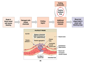
Hemostasis is the process of stopping blood loss through a series of rapid, localized reactions.
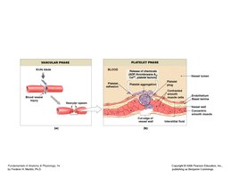
Vascular spasm: Immediate vasoconstriction after vessel injury
Platelet plug formation: Platelets adhere to exposed collagen, activate, and aggregate
Coagulation: Cascade of enzymatic reactions leading to fibrin clot formation
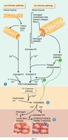
Coagulation Pathways
Coagulation involves intrinsic and extrinsic pathways, both converging to form a stable fibrin clot.
Intrinsic pathway: Initiated within blood
Extrinsic pathway: Triggered by tissue damage
Common pathway: Formation of prothrombin activator, conversion of prothrombin to thrombin, and fibrinogen to fibrin
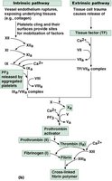
Clot Retraction and Fibrinolysis
After clot formation, platelets contract to strengthen the clot and pull vessel edges together. Fibrinolysis dissolves the clot once healing is complete.
Clot retraction: Occurs within 30–60 minutes
Fibrinolysis: Plasmin digests fibrin, restoring normal blood flow
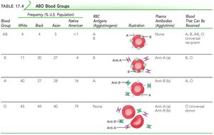
Blood Types and Transfusion
ABO and Rh Blood Groups
Blood type is determined by antigens (agglutinogens) on RBC membranes. The ABO system includes A, B, AB, and O types; the Rh system is based on the D antigen.
Type A: A antigen, anti-B antibody
Type B: B antigen, anti-A antibody
Type AB: A and B antigens, no antibodies
Type O: No antigens, anti-A and anti-B antibodies
Rh+: D antigen present
Rh-: D antigen absent
Transfusion Reactions
Transfusion reactions occur when mismatched blood is transfused, leading to agglutination and hemolysis. This can cause decreased oxygen delivery, kidney damage, and acute renal failure.
Hemolytic Disease of the Newborn (HDN)
HDN occurs when an Rh-negative mother becomes sensitized to Rh-positive fetal blood, producing antibodies that attack fetal RBCs in subsequent pregnancies. Prevention involves administration of RhoGAM.
Developmental Aspects
Blood Cell Formation Before Birth
Hematopoiesis occurs in the yolk sac, liver, and spleen before birth, then shifts to red bone marrow. Fetal hemoglobin (HbF) has a higher affinity for oxygen than adult hemoglobin (HbA).
Age-Related Blood Disorders
Blood disorders increase with age due to changes in the cardiovascular and immune systems, including higher risk of leukemia, thrombus, and embolus formation.
